Thoracic Cavity Drawing
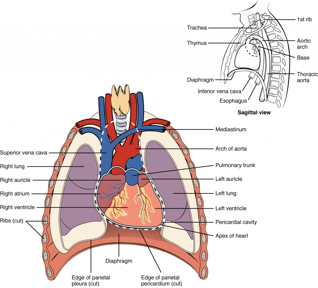
Thoracic Cavity Drawing - The chest cavity is bound by the thoracic vertebrae, which connect to the ribs that surround the cavity. Each lung is surrounded by a pleural cavity, which is formed by the visceral and parietal pleura. Web your thoracic cavity is located in your chest. The latter comprises of the first thoracic vertebrae, upper border of manubrium sterni and the first ribs on either side. An aggregate of similar cells and cell products forming a. It’s enclosed by the bones and muscles that make up your chest wall. Labeled diagrams, definitions, and lateral views included! Relating to the superior ventral cavity; Web the pathway towards the lungs is provided by airways and together, these components form the respiratory system, which is located inside the thoracic or chest cavity. Here’s a bit more detail about your thoracic cavity’s boundaries at different locations within your chest: These elements are derived from paraxial mesoderm, which forms somites from which the sclerotome, myotome, and dermatome separate. Save time with a video! Division of the anterior (ventral) cavity that houses the heart, lungs, esophagus, and trachea. The walls surrounding the thoracic cavity are made from layers of muscles and fascia that are reinforced by the thoracic skeleton (“rib cage”).. The thoracic cavity, also called the chest cavity, is a cavity of vertebrates bounded by the rib cage on the sides and top, and the diaphragm on the bottom. Web the knowledge of the sectional anatomy of the thoracic cavity is of great importance for the understanding of respective pathologies, congenital anomalies, surgery, and radiological images such as computerized. 152. The thoracic wall forms part of the axial skeleton and is composed of segmental bone, muscle, and connective tissue. The first nine ribs curve around the lateral thoracic wall and connect to the manubrium and sternum. Body cavities along with their organs and membranes simplified! Narrower above and wider below. It’s enclosed by the bones and muscles that make up. Web choose from thoracic cavity stock illustrations from istock. Subscribe subscribed unsubscribe embed share report collection. Narrower above and wider below. Web this thoracic and pulmonary anatomy tool is especially designed for students of anatomy (medical and paramedical studies). Web the thoracic wall consists of a bony framework held together by twelve thoracic vertebrae posteriorly, giving rise to ribs that. Web how to draw a thoracic cavity (front view) mi's creation studio. Web your thoracic cavity is located in your chest. Web the thoracic wall consists of a bony framework held together by twelve thoracic vertebrae posteriorly, giving rise to ribs that encircle the lateral and anterior thoracic cavity. The chest cavity is bound by the thoracic vertebrae, which connect. Web your thoracic cavity is located in your chest. These triangles should be angled upward at the base in the medial direction. Web 3d interactive modules and video tutorials on the anatomy of the thoracic cavity, including the heart, lungs, breast, chest wall, and respiratory tract. Web thoracic cavity, the second largest hollow space of the body. Web the thoracic. The walls surrounding the thoracic cavity are made from layers of muscles and fascia that are reinforced by the thoracic skeleton (“rib cage”). The first nine ribs curve around the lateral thoracic wall and connect to the manubrium and sternum. Web your thoracic cavity is located in your chest. It is enclosed by the ribs, the vertebral column, and the. Web the thoracic wall consists of a bony framework held together by twelve thoracic vertebrae posteriorly, giving rise to ribs that encircle the lateral and anterior thoracic cavity. Among the major organs contained in the thoracic cavity are the heart and lungs. Web the thoracic cavity communicates with the neck via the superior thoracic aperture and with the abdominal cavity. Your thoracic cavity begins just below your neck and ends at the bottom of your ribcage. Web how to draw a thoracic cavity (front view) mi's creation studio. It is enclosed by the ribs, the vertebral column, and the sternum, or breastbone, and is separated from the abdominal cavity by the diaphragm. It’s enclosed by the bones and muscles that. Save time with a video! The lungs lie either side of the mediastinum, within the thoracic cavity. Web 3d interactive modules and video tutorials on the anatomy of the thoracic cavity, including the heart, lungs, breast, chest wall, and respiratory tract. Web this thoracic and pulmonary anatomy tool is especially designed for students of anatomy (medical and paramedical studies). Web. Among the major organs contained in the thoracic cavity are the heart and lungs. The walls surrounding the thoracic cavity are made from layers of muscles and fascia that are reinforced by the thoracic skeleton (“rib cage”). The chest cavity is bound by the thoracic vertebrae, which connect to the ribs that surround the cavity. Subscribe subscribed unsubscribe embed share report collection. Web 3d interactive modules and video tutorials on the anatomy of the thoracic cavity, including the heart, lungs, breast, chest wall, and respiratory tract. Anatomical illustrations of the lungs. The first nine ribs curve around the lateral thoracic wall and connect to the manubrium and sternum. Web the thoracic cavity or the chest cavity lies between the neck and the abdomen. 152 views 1 year ago daily drawing practice. The latter comprises of the first thoracic vertebrae, upper border of manubrium sterni and the first ribs on either side. These elements are derived from paraxial mesoderm, which forms somites from which the sclerotome, myotome, and dermatome separate. Web the knowledge of the sectional anatomy of the thoracic cavity is of great importance for the understanding of respective pathologies, congenital anomalies, surgery, and radiological images such as computerized. Undergoes contraction and relaxation, altering the volume of the thoracic cavity and. Web how to draw a thoracic cavity (front view) mi's creation studio. Here’s a bit more detail about your thoracic cavity’s boundaries at different locations within your chest: Web separates the thoracic cavity from the abdominal cavity (the word diaphragm is derived from the greek ‘diáphragma’, meaning partition).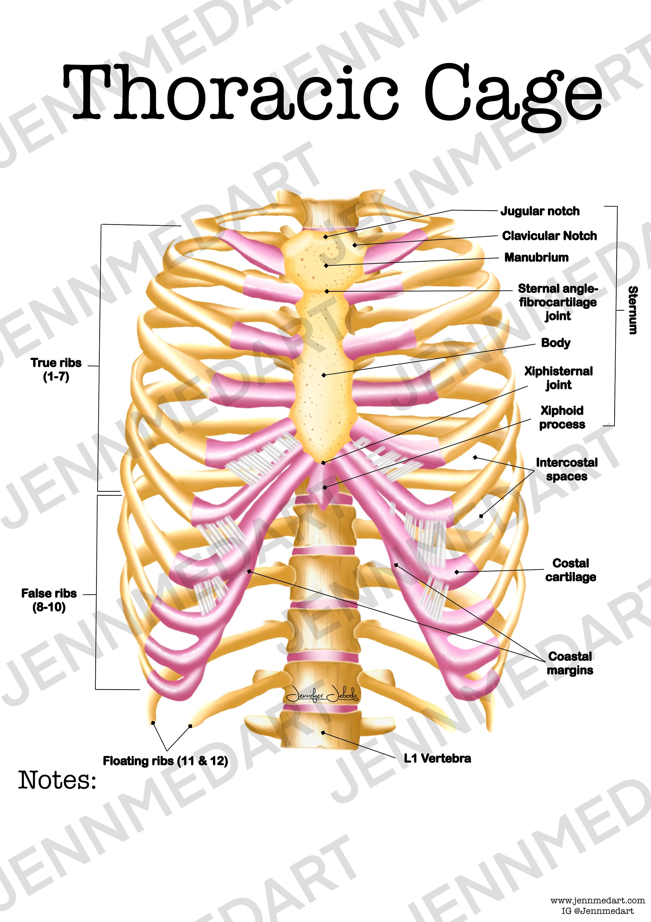
Thoracic Cage Anatomy Worksheet Single FILLED Digital Download Human

Thoracic Cavity by Cryssari on DeviantArt

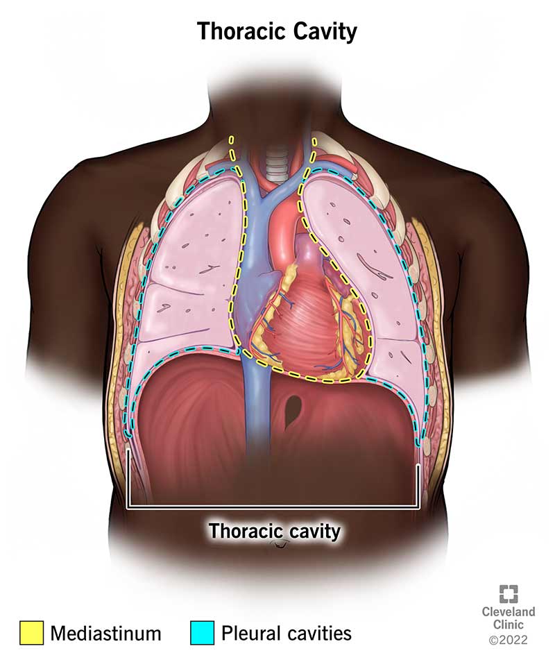
The lungs, trachea and bronchi, mediastinum and detail of chest wall
Body Cavity Diagram ClipArt Best
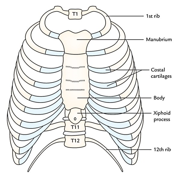
Thoracic Cage Intrinsic Muscles, Formation and Shape Earth's Lab
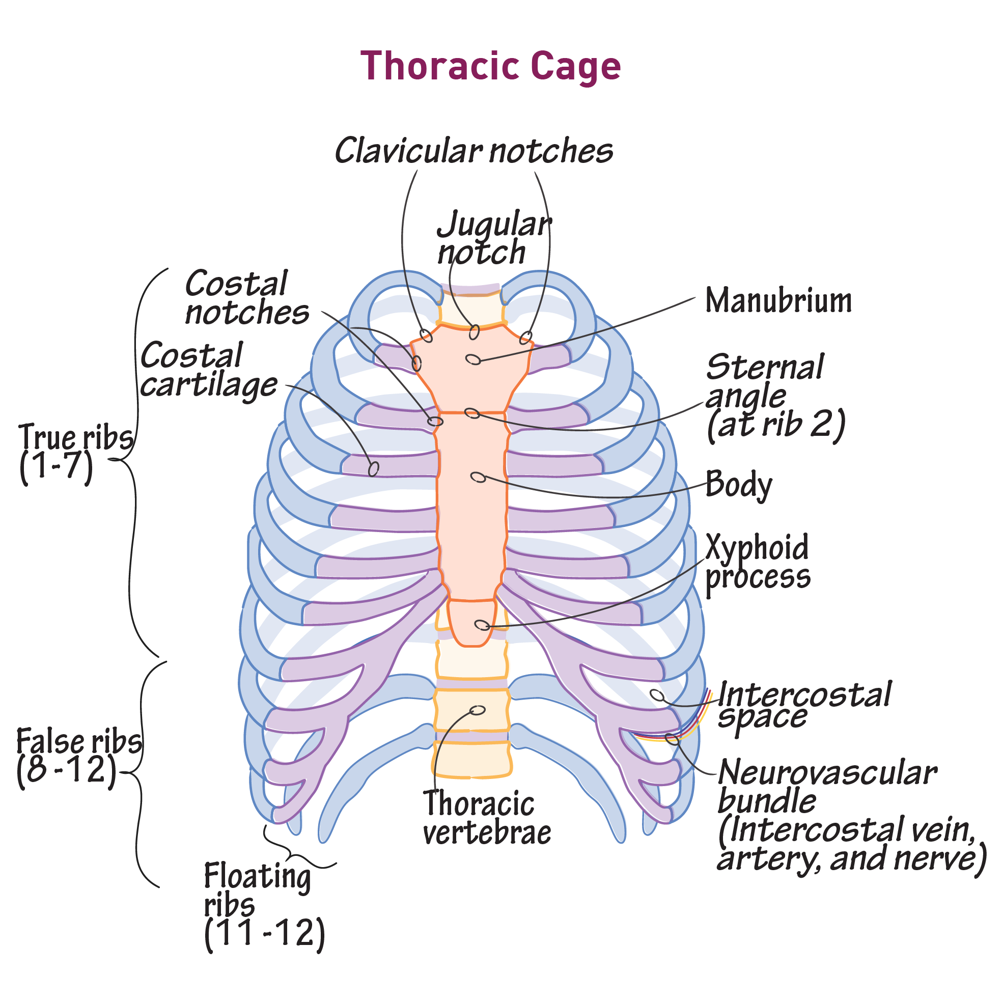
Gross Anatomy Glossary Thoracic Cage Draw It to Know It

Human Anatomy Chest Cavity Anatomy Of Chest Bones Human Anatomy Diagram
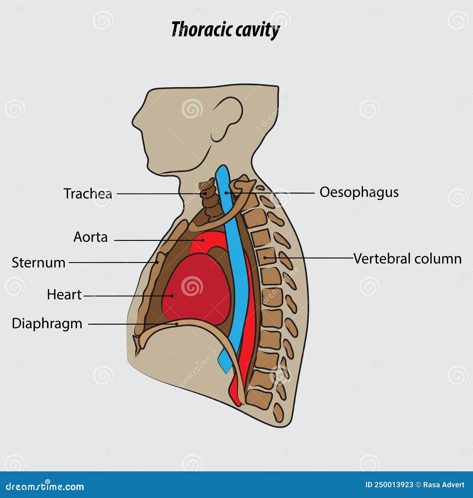
Thoracic Cavity Vector Illustration Drawing Labeled Stock Vector
Thoracic Cavity Location and Function

WHAT IS THORACIC CAVITY (ANATOMY) INFO HUB YouTube
The Thoracic Cage And Walls Enclose This Cavity And Its Structures, And Play An Essential Role In Pulmonary Ventilation.
It Is Enclosed By The Ribs, The Vertebral Column, And The Sternum, Or Breastbone, And Is Separated From The Abdominal Cavity By The Diaphragm.
Division Of The Anterior (Ventral) Cavity That Houses The Heart, Lungs, Esophagus, And Trachea.
It Is Bounded By The Thoracic Wall And Extends From The Diaphragm (Below) To The Superior Thoracic Aperture (Above).
Related Post: