Simple Squamous Drawing
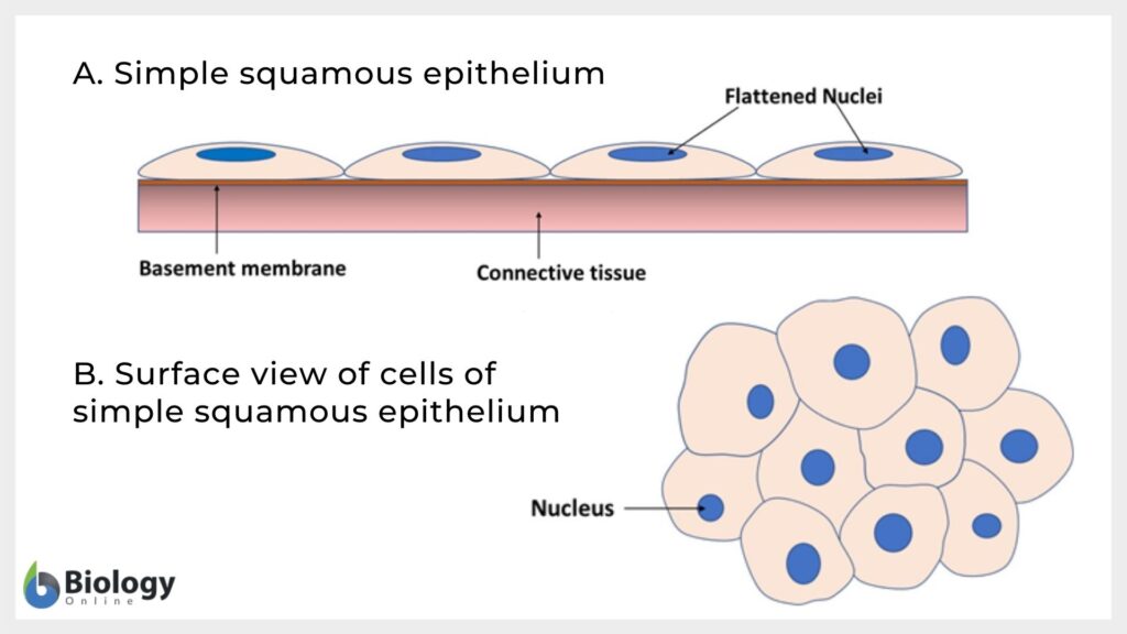
Simple Squamous Drawing - Absorption and filtration processes classes: • how to draw simple squamous epitheliu. The typical example of the simple squamous epithelium will be found in the lung’s alveoli, the parietal layer of the bowman’s capsule of the kidney, and the loop of henle of kidney tubules. Squamous cells are large, thin, and flat and contain a rounded nucleus. Like other epithelial cells, they have polarity and contain a distinct apical surface with specialized membrane proteins. Web distinguish between simple epithelia and stratified epithelia, as well as between squamous, cuboidal, and columnar epithelia. Web simple squamous epithelium can be found in many locations in the body (e.g., lining blood vessels, lining the alveoli (air sacs) of our lungs, and in bowman’s capsule of the kidney). A cuboidal epithelial cell looks close to a square. A columnar epithelial cell looks like a column or a tall rectangle. However, the focus here is only on a simple squamous epithelium. A squamous epithelial cell looks flat under a microscope. Web simple squamous epithelium is a type of simple epithelium made up of squamous epithelial cells that lines the outer layer of the skin, endothelium, and secretory parts of the small glands. Learn about its location in the body, cells, and characteristics. Notice that the location of the. Simple squamous epithelium. Web simple squamous epithelium is a type of simple epithelium made up of squamous epithelial cells that lines the outer layer of the skin, endothelium, and secretory parts of the small glands. It also lines the glomeruli in the kidney and the pulmonary alveoli where passive diffusion occurs. Absorption and filtration processes classes: What is simple squamous epithelium? Learn more. 19k views 2 years ago cell biology. Describe the structure and function of endocrine and exocrine glands. Learn more about how pressbooks supports open publishing practices. Web simple squamous epithelium is a type of simple epithelium made up of squamous epithelial cells that lines the outer layer of the skin, endothelium, and secretory parts of the small glands. Web drawing. Web drawing histological diagram of simple squamous epithelia.useful for all medical students.drawn by using h & e pencils Web simple squamous epithelium diagram | quizlet. What is simple squamous epithelium? A simple squamous epithelium is a single layer of flat. Describe the structure and function of endocrine and exocrine glands. Blood and lymphatic vessels, air sacs of lungs, lining of the heart Web key facts about the simple epithelium; Web distinguish between simple epithelia and stratified epithelia, as well as between squamous, cuboidal, and columnar epithelia. However, the focus here is only on a simple squamous epithelium. Web histology diagram of simple squamous epithelium histology diagram. Web drawing histological diagram of simple squamous epithelia.useful for all medical students.drawn by using h & e pencils The typical example of the simple squamous epithelium will be found in the lung’s alveoli, the parietal layer of the bowman’s capsule of the kidney, and the loop of henle of kidney tubules. Learn more about how pressbooks supports open publishing practices.. Web a simple epithelium is one cell layer thick, and the cells may be squamous, cuboidal, or columnar in shape. 19k views 2 years ago cell biology. The shape of the cells in the single cell layer of simple epithelium reflects the functioning of those cells. Squamous cells are large, thin, and flat and contain a rounded nucleus. To help. A columnar epithelial cell looks like a column or a tall rectangle. Learn more about how pressbooks supports open publishing practices. Squamous cells are large, thin, and flat and contain a rounded nucleus. 19k views 2 years ago cell biology. Author keta bhakta view bio. The cells in simple squamous epithelium have the appearance of thin scales. A simple squamous epithelium is a single layer of flat. • how to draw simple squamous epitheliu. Web key facts about the simple epithelium; Terms in this set (4) location. Web key facts about the simple epithelium; Absorption and filtration processes classes: Author keta bhakta view bio. Terms in this set (4) location. Web there are three basic shapes used to classify epithelial cells. Like other epithelial cells, they have polarity and contain a distinct apical surface with specialized membrane proteins. To help you understand how to identify simple squamous epithelium, we have included two examples of this tissue. Learn more about how pressbooks supports open publishing practices. Each contains a glomerulus (a tuft of capillaries) surrounded by bowman's capsule. A squamous epithelial cell looks flat under a microscope. Find one of the round structures (~250 µm diameter) known as renal corpuscles. Describe the structure and function of endocrine and exocrine glands. It also lines the glomeruli in the kidney and the pulmonary alveoli where passive diffusion occurs. Web distinguish between simple epithelia and stratified epithelia, as well as between squamous, cuboidal, and columnar epithelia. What is simple squamous epithelium? The cells in simple squamous epithelium have the appearance of thin scales. Web want to create or adapt books like this? Learn about its location in the body, cells, and characteristics. Terms in this set (4) location. A columnar epithelial cell looks like a column or a tall rectangle. Web simple squamous epithelium diagram | quizlet.
Simple Squamous Epithelium Function Location Structure And Histology

Simple Squamous Epithelium Diagram Quizlet

34+ Simple Squamous Epithelium Drawing NeeraNatania
![[1/7] A simple squamous epithelium drawing](https://preview.redd.it/ogmt2h7pgop11.jpg?width=960&crop=smart&auto=webp&s=924d5c709e144fe9344374f66ac7e64073cf12e1)
[1/7] A simple squamous epithelium drawing
Simple Squamous Epithelium Function Location Structure

Cuboidal Epithelial Tissue
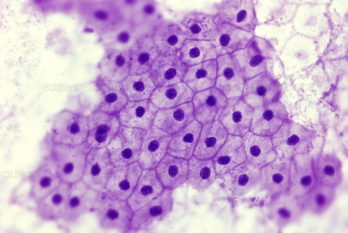
Simple Squamous Epithelial Tissue Under Microscope
Simple Squamous Epithelium Labeled
Simple Squamous Epithelium Inrtroducrion , Anatomy & Function
Simple Cuboidal Epithelium Labeled Basement Membrane
Web Simple Squamous Epithelium Can Be Found In Many Locations In The Body (E.g., Lining Blood Vessels, Lining The Alveoli (Air Sacs) Of Our Lungs, And In Bowman’s Capsule Of The Kidney).
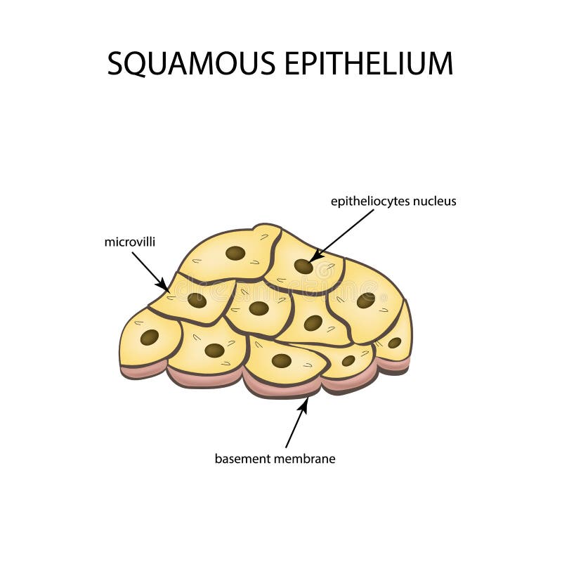
Squamous Cells Are Large, Thin, And Flat And Contain A Rounded Nucleus.
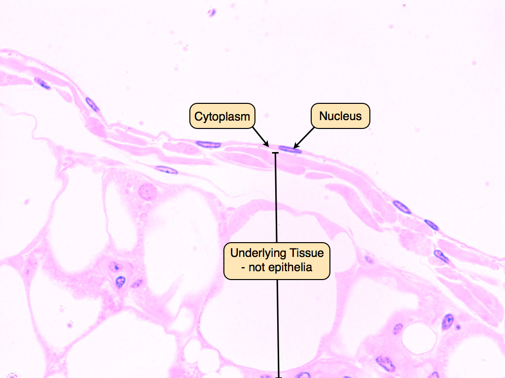
Web Simple Squamous Epithelia Are Tissues Formed From One Layer Of Squamous Cells That Line Surfaces.
Web Drawing Histological Diagram Of Simple Squamous Epithelia.useful For All Medical Students.drawn By Using H & E Pencils
Related Post: