Paramecium Drawing
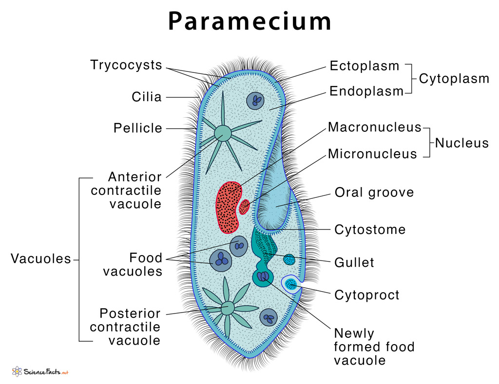
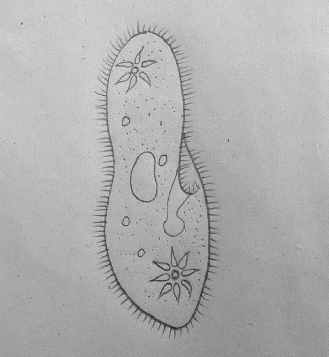
Paramecium Drawing - Web © 2023 google llc. It ranges from 50 to 300um in size which varies from species to species. Paramecium is a ciliate protozoan. They are characterised by the presence of thousands of cilia covering their body. Paramecium structure consists of trichocysts, contractile vacuoles, and cilia among other specialized organelles. Uniform ciliation all over body except at post, end where ciliation are large & form a caudal tuft. Light microscopic appearance of paramecium caudatum. How does a paramecium eat? Web panel 1 (a,b): At the end of the gullet, food vacuoles are formed. Web © 2023 google llc. Drawing of paramecium illustrating light microscopic features: It is mostly found in a freshwater environment. Web i am from gurgaon. The area of the paramecium appears pinched inward and is called the oral groove, cilia sweep food into this area. It is very easy drawing detailed method to help you. Uniform ciliation all over body except at post, end where ciliation are large & form a caudal tuft. Web this will also help you to draw the structure and diagram of paramecium. Web i am from gurgaon. Web hello friends in this video i will tell you about how to. It ranges from 50 to 300um in size which varies from species to species. Web this will also help you to draw the structure and diagram of paramecium. Web the structure of pellicle and cilia. Paramecium is a ciliate protozoan. Uniform ciliation all over body except at post, end where ciliation are large & form a caudal tuft. They are found in freshwater, marine and brackish water. 2.2k views 10 months ago easy science drawing. The specialized “skin” of paramecium cell body. Paramecia, illustrated by otto müller, 1773. How fast can a paramecium move? It is very easy drawing detailed method to help you. In this video, i will be showing you how to draw and color a paramecium diagram easily for kids and others. Web species of paramecium vary widely in size from 50 to 330 µm (0.0020 to 0.0130 in) and thus can be viewed under a light microscope. A paramecium is. #paramecium #howtodraw #biology this is a diagram of the paramecium. Fresh water, free living, omnipresent and is found in stagnant water. Drawing of paramecium illustrating light microscopic features: They are characterised by the presence of thousands of cilia covering their body. Cytostome, cytopharynx, and food vacuole. Web © 2023 google llc. Unlike amoeba, paramecium has a distinct and permanent shape and certain areas of cytoplasm, (cell organelles), are specialised to carry out specific functions. Web panel 1 (a,b): Fresh water, free living, omnipresent and is found in stagnant water. How to draw paramecium step by step/paramecium drawing easy. What is inside the cell. Web paramecium is a unicellular organism with a shape resembling the sole of a shoe. Fresh water, free living, omnipresent and is found in stagnant water. 25k views 2 years ago #howtodraw #biology #paramecium. Web this will also help you to draw the structure and diagram of paramecium. Drawing of paramecium illustrating light microscopic features: #paramecium #howtodraw #biology this is a diagram of the paramecium. Paramecium structure consists of trichocysts, contractile vacuoles, and cilia among other specialized organelles. Download 77 paramecium diagram stock illustrations, vectors & clipart for free or amazingly low rates! Food vacuoles then remain in the cytoplasm until the food is digested. Does a paramecium make a poo? Cv contractile vacuoles, fv food vacuoles, manu macronucleus, mino micronucleus, pe peristome, tr trichocysts and ve vestibulum. Web paramecium is a unicellular organism with a shape resembling the sole of a shoe. Sience biology in easy steps and compact way. 25k views 2 years ago #howtodraw #biology #paramecium. What is inside the cell. The basic anatomy of paramecium shows the following distinct and specialized. Web i am from gurgaon. Cv contractile vacuoles, fv food vacuoles, manu macronucleus, mino micronucleus, pe peristome, tr trichocysts and ve vestibulum. Light microscopic appearance of paramecium caudatum. Web published 21 february 2022. Web food enters the paramecium through the mouth pore (color orange) and goes to the gullet (color dark blue). They are also found attached to the surface. Paramecium is a ciliate protozoan. #paramecium #howtodraw #biology this is a diagram of the paramecium. 151k views 3 years ago science diagrams | explained and labelled science diagrams. How to draw paramecium step by step/paramecium drawing easy. A paramecium is a microscopic organism that lives in ponds and streams. 25k views 2 years ago #howtodraw #biology #paramecium. It ranges from 50 to 300um in size which varies from species to species. See how cilia do the wave.
Paramecium Definition, Structure, Characteristics and Diagram

Paramecium stock illustration. Image of outline, anatomy 33357022

How TO Draw paramecium easy with pencil YouTube

Simplest Way Of Drawing Paramecium Diagram How To Draw Paramecium in

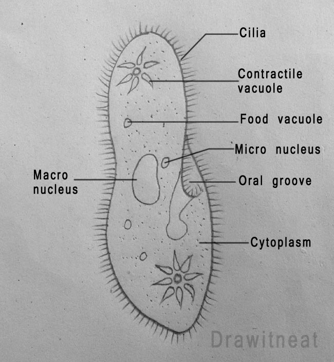
DRAW IT NEAT How to draw Paramecium

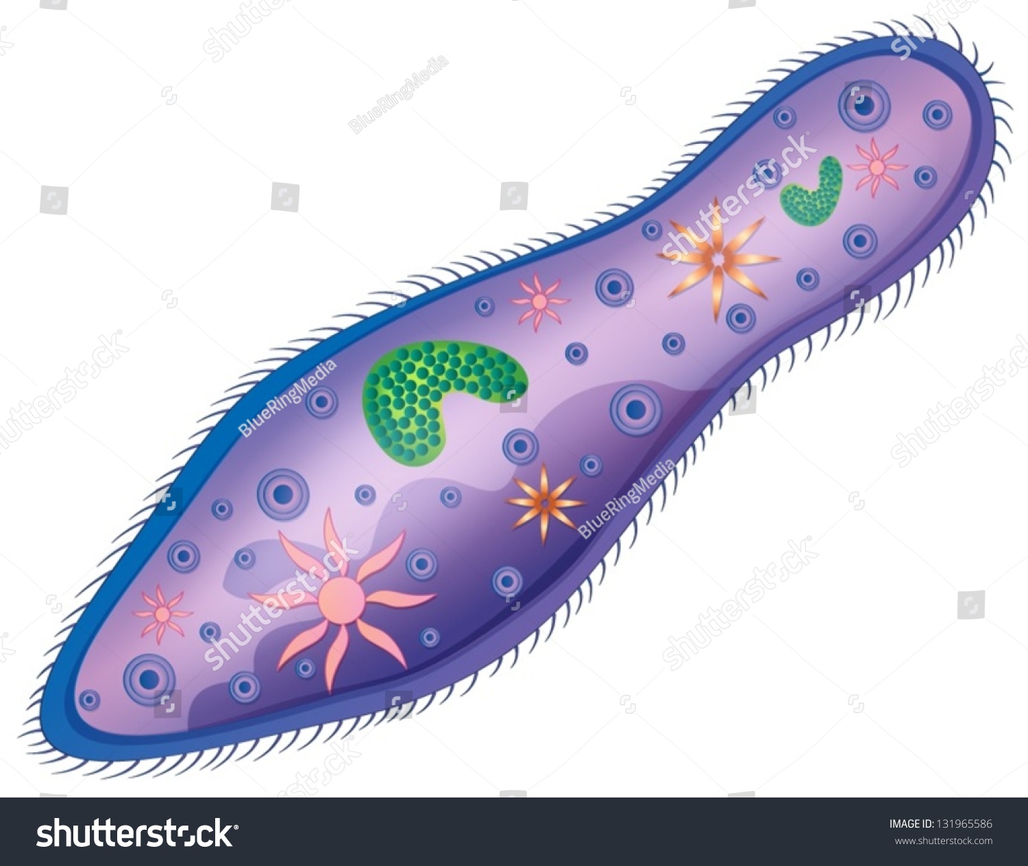
Sketch Of A Paramecium Stock Vector Illustration 131965586 Shutterstock

Paramecium diagram by lucidhysteria on DeviantArt

Paramecium in Colour and Doodle on White Background Stock Vector

DRAW IT NEAT How to draw Paramecium

How to draw paramecium step by step easy paramecium diagram YouTube
Food Vacuoles Then Remain In The Cytoplasm Until The Food Is Digested.
Web Species Of Paramecium Vary Widely In Size From 50 To 330 Μm (0.0020 To 0.0130 In) And Thus Can Be Viewed Under A Light Microscope.
The Area Of The Paramecium Appears Pinched Inward And Is Called The Oral Groove, Cilia Sweep Food Into This Area.
They Are Characterised By The Presence Of Thousands Of Cilia Covering Their Body.
Related Post: