Larynx Drawing
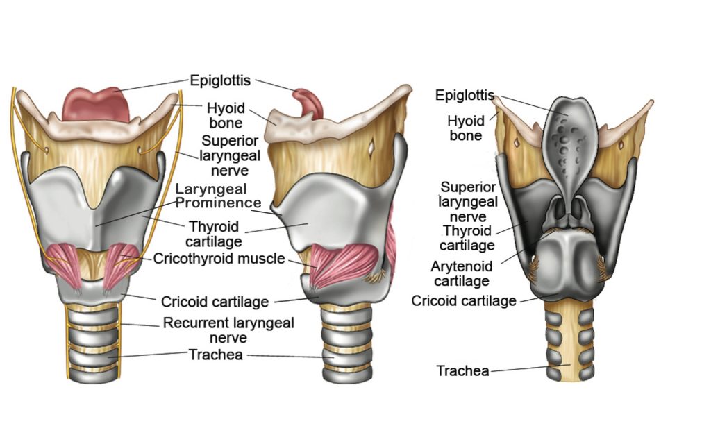
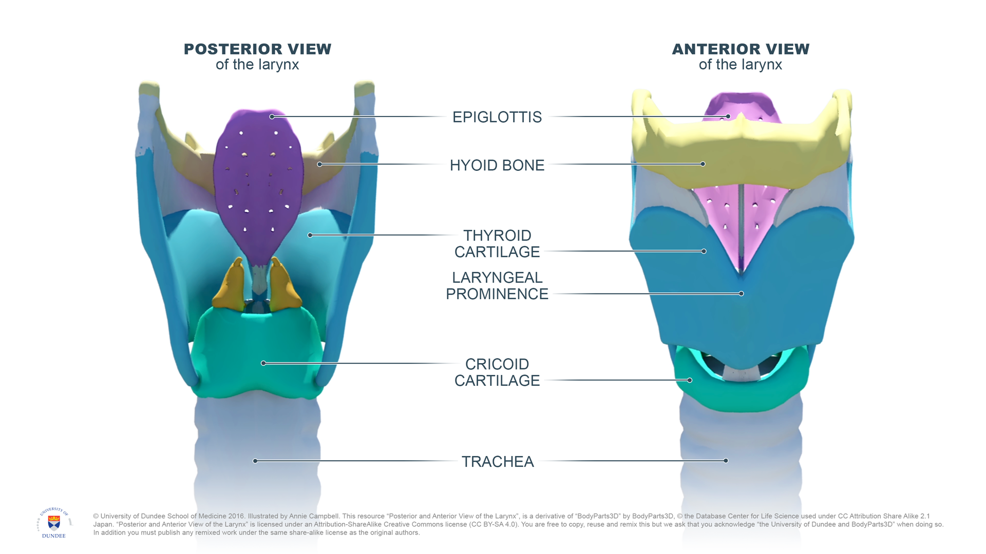
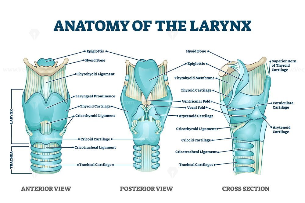
Larynx Drawing - Medically reviewed by isabel casimiro, md, phd. Drawing shows the epiglottis, supraglottis, glottis, subglottis, and vocal cords. The three parts of the larynx are the supraglottis (including the epiglottis), the glottis (including the vocal cords), and the subglottis. Transverse and oblique arytenoid muscles. It’s a hollow tube that’s about 4 to 5 centimeters (cm) in length and width. Larynx, a hollow, tubular structure connected to the top of the windpipe (trachea); The anatomy of the larynx. Also shown are the tongue, trachea, and esophagus. The larynx ( / ˈlærɪŋks / ), commonly called the voice box, is an organ in the top of the neck involved in breathing, producing sound and protecting the trachea against food aspiration. Web what is larynx (voice box) definition, where is it located, anatomy (cartilages, muscles, innervations), what does the larynx do, picture, diagram. It surrounds and protects the vocal chords, as well as the entrance to the trachea, preventing food particles or fluids from entering the lungs. It is situated between the trachea and the root of the tongue, at the upper and forepart of the neck, where it presents a considerable projection in the middle line. Web the larynx is the most. Web the larynx is the most superior part of the respiratory tract in the neck and the voice box of the human body. Larynx, a hollow, tubular structure connected to the top of the windpipe (trachea); 3d anatomy tutorial on the cartilages of the larynx from anatomyzone for more videos, 3d models and notes visit:. It consists of three sections:.. Used as teaching material, this model depicts the muscles, cartilages and ligaments of the larynx. Drawing shows the epiglottis, supraglottis, glottis, subglottis, and vocal cords. How to draw larynx | how to draw larynx step by step | how to draw larynx class 8 | larynxhello friends in this video i tell you about how can we draw. 3d anatomy. It consists of three sections:. It is situated between the trachea and the root of the tongue, at the upper and forepart of the neck, where it presents a considerable projection in the middle line. Also shown are the tongue, trachea, and esophagus. Web cartilaginous framework and ligaments. Arteries and nerves and b. The walls of the larynx are made up of cartilage, ligaments, membranes, muscles, and respiratory mucosa (or mucous. The larynx is composed of three large unpaired cartilages (cricoid, thyroid, and epiglottis) and three paired smaller cartilages (arytenoid, corniculate, and cuneiform), making a total of nine individual cartilages. Your larynx (voice box) helps you to breathe. Also shown are the tongue,. This was modelled utlising ct data, imported via invesalius 3.1, and original sculpting in pixologic zbrush. Also shown are the tongue, trachea, and esophagus. Web explore the anatomy and function of the larynx, the voice box that connects the pharynx and the trachea. Arteries and nerves and b. Medically reviewed by isabel casimiro, md, phd. This was modelled utlising ct data, imported via invesalius 3.1, and original sculpting in pixologic zbrush. Arteries and nerves and b. The cartilages of the larynx make up its skeleton. The larynx is composed of three large unpaired cartilages (cricoid, thyroid, and epiglottis) and three paired smaller cartilages (arytenoid, corniculate, and cuneiform), making a total of nine individual cartilages. Medically. The extrinsic muscles of the larynx are those that are somehow attached to the hyoid bone, be it via origin or insertion and thus move the thyroid cartilage. Neurovasculature of the larynx and trachea a. This was modelled utlising ct data, imported via invesalius 3.1, and original sculpting in pixologic zbrush. It contains your vocal cords, so you can make. Web what is larynx (voice box) definition, where is it located, anatomy (cartilages, muscles, innervations), what does the larynx do, picture, diagram. Cartilaginous skeleton of the larynx and trachea a. Web explore the anatomy and function of the larynx, the voice box that connects the pharynx and the trachea. The primary function of the larynx in humans and other vertebrates. Find out how it works, its disorders and clinical relevance. Also shown are the tongue, trachea, and esophagus. Web explore the anatomy and function of the larynx, the voice box that connects the pharynx and the trachea. The three parts of the larynx are the supraglottis (including the epiglottis), the glottis (including the vocal cords), and the subglottis. Transverse and. The walls of the larynx are made up of cartilage, ligaments, membranes, muscles, and respiratory mucosa (or mucous. Web cartilaginous framework and ligaments. The larynx is composed of three large unpaired cartilages (cricoid, thyroid, and epiglottis) and three paired smaller cartilages (arytenoid, corniculate, and cuneiform), making a total of nine individual cartilages. Also shown are the tongue, trachea, and esophagus. The extrinsic muscles of the larynx are those that are somehow attached to the hyoid bone, be it via origin or insertion and thus move the thyroid cartilage. Larynx, a hollow, tubular structure connected to the top of the windpipe (trachea); The anatomy of the larynx. The cartilages of the larynx make up its skeleton. Your larynx is part of your respiratory system. Your larynx (voice box) helps you to breathe. 1.2m views 11 years ago respiratory system. Also shown are the tongue, trachea, and esophagus. Web how to draw larynx easily/human larynx diagram.it is very easy drawing detailed method to help you.i draw the human larynx with pencil on art paper on my ea. Drawing shows the epiglottis, supraglottis, glottis, subglottis, and vocal cords. Neurovasculature of the larynx and trachea a. Transverse and oblique arytenoid muscles.
Larynx, drawing Stock Image C006/3934 Science Photo Library

Larynx structure, function, cartilages, muscles, blood supply and vocal

Medical Images Art & Science Graphics

Larynx, Drawing Stock Image C024/4350 Science Photo Library

Larynx Anatomy Concise Medical Knowledge
/human-larynx--illustration-1190674300-4ce616b410ea488ab61b6fca58fc992b.jpg)
Larynx Anatomy, Function, and Treatment

Dundee Drawing Posterior and Anterior View of the Larynx English

Larynx anatomy with labeled structure scheme and educational medical

16. The Larynx SimpleMed Learning Medicine, Simplified

Diagram Of Larynx With Labeling
Web What Is Larynx (Voice Box) Definition, Where Is It Located, Anatomy (Cartilages, Muscles, Innervations), What Does The Larynx Do, Picture, Diagram.
Arteries And Nerves And B.
What Is The Larynx (Voice Box)?
Web Explore The Anatomy And Function Of The Larynx, The Voice Box That Connects The Pharynx And The Trachea.
Related Post: