Hyaline Cartilage Tissue Drawing
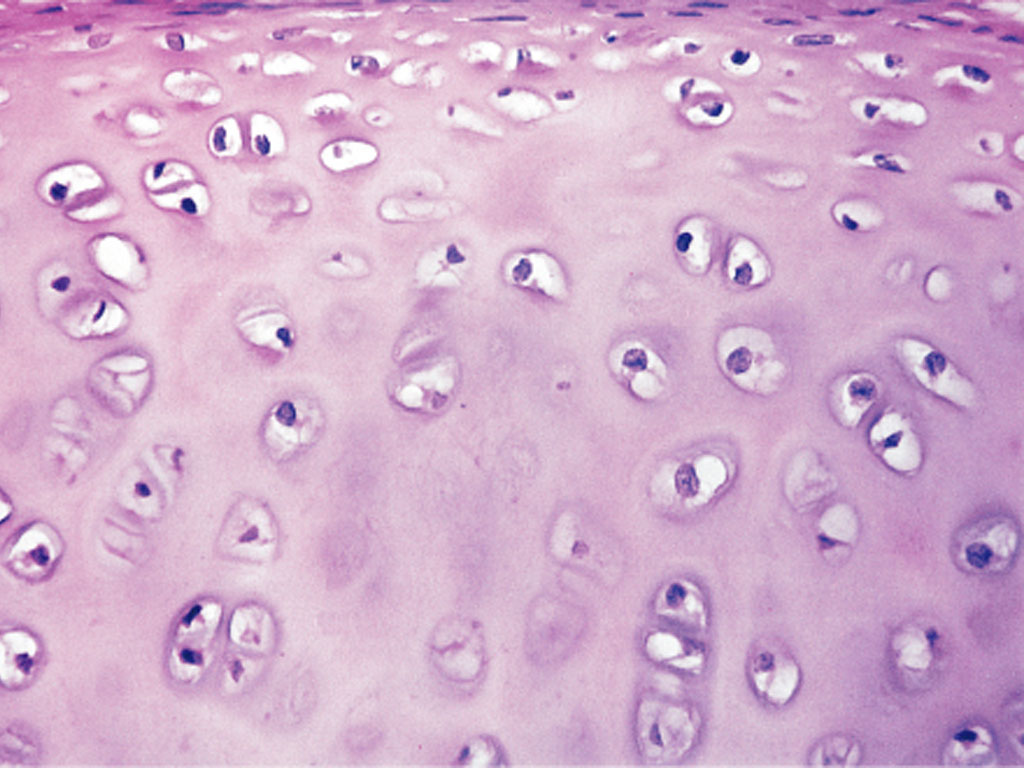
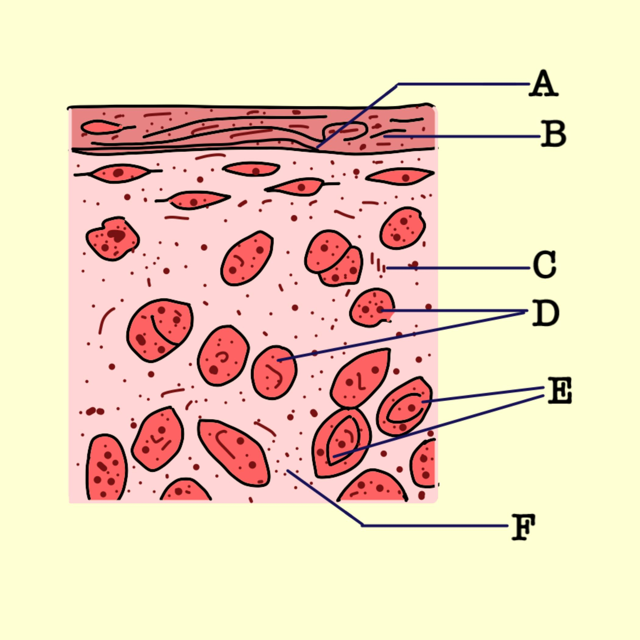
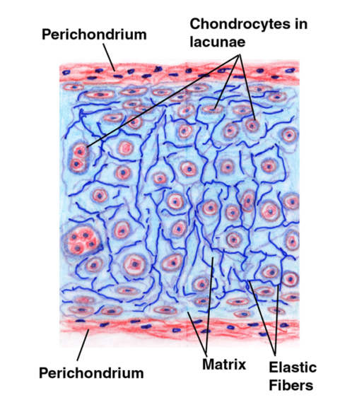
Hyaline Cartilage Tissue Drawing - Web the hyaline cartilage in the trachea is in the middle of the tracheal wall. Isogenous groups and interstitial growth results when chondrocytes divide and produce extracellular matrix. Web during embryonic development, hyaline cartilage serves as temporary cartilage models that are essential precursors to the formation of most of the axial and. Watch the video tutorial now. It is avascular and its microarchitecture is less organized than bone. 35 views 4 months ago histology discussion, viva, oral questions. Learn more about how pressbooks supports open publishing practices. It is typically characterized by a firm consistency and a smooth surface. Web hyaline cartilage, the most common type of cartilage, is composed of type ii collagen and chondromucoprotein and often has a glassy appearance. Territorial matrix lies immediately around each. Territorial matrix lies immediately around each. Most of the image is occupied by a section. A histological overview of the most common type of cartilage in the human body. Web during embryonic development, hyaline cartilage serves as temporary cartilage models that are essential precursors to the formation of most of the axial and. It is avascular and its microarchitecture is. 35 views 4 months ago histology discussion, viva, oral questions. Tamás oláh, tunku kamarul, henning madry & malliga raman murali. Isogenous groups and interstitial growth results when chondrocytes divide and produce extracellular matrix. Web during embryonic development, hyaline cartilage serves as temporary cartilage models that are essential precursors to the formation of most of the axial and. Hyaline cartilage with. Tamás oláh, tunku kamarul, henning madry & malliga raman murali. Web likecomment share subscribe #hyalinecartilage #histodiagrams #hyalinecartilagediagram #cartilagehistology Web hyaline cartilage is the most common type of cartilage in the human body. Cartilage is a flexible connective tissue that differs from bone in several ways; Web articular cartilage is a remnant of the hyaline cartilage that formed the template for. Web want to create or adapt books like this? Territorial matrix lies immediately around each. It tends to stain more blue than other kinds of connective tissue (however, remember that color should. Although hyaline cartilage feels nearly as hard and dense as bone. Web the hyaline cartilage in the trachea is in the middle of the tracheal wall. Check out our youtube video to help you understand hyaline cartilage: Hyaline cartilage with and without illustration overlay. A higher magnification of the wall of the trachea shows the lumen with its epithelial lining in the lower left of the image. A histological overview of the most common type of cartilage in the human body. Tamás oláh, tunku kamarul, henning. Most of the image is occupied by a section. Cartilage is a flexible connective tissue that differs from bone in several ways; Watch the video tutorial now. It tends to stain more blue than other kinds of connective tissue (however, remember that color should. Territorial matrix lies immediately around each. Web articular cartilage is a remnant of the hyaline cartilage that formed the template for the developing bone. Learn more about how pressbooks supports open publishing practices. Check out our youtube video to help you understand hyaline cartilage: Isogenous groups and interstitial growth results when chondrocytes divide and produce extracellular matrix. Web during embryonic development, hyaline cartilage serves as temporary. Although hyaline cartilage feels nearly as hard and dense as bone. Hyaline cartilage with and without illustration overlay. It tends to stain more blue than other kinds of connective tissue (however, remember that color should. Learn more about how pressbooks supports open publishing practices. Web the illustrative book of cartilage repair. It is typically characterized by a firm consistency and a smooth surface. It is avascular and its microarchitecture is less organized than bone. Web hyaline cartilage, the most common type of cartilage, is composed of type ii collagen and chondromucoprotein and often has a glassy appearance. Watch the video tutorial now. Hyaline cartilage with and without illustration overlay. Check out our youtube video to help you understand hyaline cartilage: Although hyaline cartilage feels nearly as hard and dense as bone. New articular cartilage is limited to interstitial growth because of the. Web hyaline cartilage, the most common type of cartilage, is composed of type ii collagen and chondromucoprotein and often has a glassy appearance. Slide 2, trachea (h&e). Territorial matrix lies immediately around each. Isogenous groups and interstitial growth results when chondrocytes divide and produce extracellular matrix. Web hyaline cartilage, the most common type of cartilage, is composed of type ii collagen and chondromucoprotein and often has a glassy appearance. It tends to stain more blue than other kinds of connective tissue (however, remember that color should. The lack of blood vessels in hyaline cartilage means that nutrients and. Web the hyaline cartilage in the trachea is in the middle of the tracheal wall. Web during embryonic development, hyaline cartilage serves as temporary cartilage models that are essential precursors to the formation of most of the axial and. Cartilage is a flexible connective tissue that differs from bone in several ways; It is avascular and its microarchitecture is less organized than bone. Hyaline cartilage with and without illustration overlay. A higher magnification of the wall of the trachea shows the lumen with its epithelial lining in the lower left of the image. Web likecomment share subscribe #hyalinecartilage #histodiagrams #hyalinecartilagediagram #cartilagehistology Check out our youtube video to help you understand hyaline cartilage: Tamás oláh, tunku kamarul, henning madry & malliga raman murali. Web hyaline cartilage is a supportive connective tissue with a rigid yet slightly flexible extracellular matrix. Web articular cartilage is a remnant of the hyaline cartilage that formed the template for the developing bone.
Mammal. Hyaline cartilage. Transverse section. 250X Hyaline cartilage
Hyaline Cartilage
Hyaline Cartilage Connective Tissue Labeled

How to Draw Hyaline Cartilage Simple and easy steps Biology Exam

Perichondrium as hyaline, fibrous and elastic cartilage membrane

Illustrations Hyaline Cartilage General Histology

Hyaline cartilage structure and biochemical composition. Schematic

Connective Tissue Anatomy and Physiology
Hyaline cartilage Definition and Examples Biology Online Dictionary

Histology Image Cartilage
Web Want To Create Or Adapt Books Like This?
Learn More About How Pressbooks Supports Open Publishing Practices.
Although Hyaline Cartilage Feels Nearly As Hard And Dense As Bone.
A Histological Overview Of The Most Common Type Of Cartilage In The Human Body.
Related Post: