Human Cell Drawing
Human Cell Drawing - View human cell drawing videos. Number 1 shows the nucleus, numbers 3 to 13 show different organelles immersed in the cytosol, and number 14 on the surface of the cell shows the plasma membrane. Web the human cell diagram unlabeled is a visual representation of the different parts of a typical human cell without any labels or descriptions. Web browse over 50,000 icons and templates from over 30 fields of life science, curated and vetted by industry professionals. Immune system human anatomy, blood cell or. All cells have a cell membrane that separates the inside and the outside of the cell, and controls what goes in and comes out. Place another circle in the middle of it to show the nucleus. Web interactive guide to stem cells and cell biology with 3d models and real microscopy data of gfp labeled hipscs. Skin tissue cells, layers of skin, blood in vein. Low poly wireframe human cell or embryonic stem cell microscope background. The cell membrane is the outer coating of the cell and contains the cytoplasm, substances within it and the organelle. View human cell diagram videos. Web the synthetic cells were stable even at 122 degrees fahrenheit, opening up the possibility of manufacturing cells with extraordinary capabilities in environments normally unsuitable to human life. Web the synthetic cells were stable even. Within the nucleus is a spherical body known as the nucleolus, which contains clusters of protein, dna, and rna. The top half of the cell volume was removed. All cells have a cell membrane that separates the inside and the outside of the cell, and controls what goes in and comes out. Vector illustration of the animal cell anatomy structure.. Most cells have only one nucleus, but some have more than one, and others—like mature red blood cells—don’t have one at all. Vector illustration of the animal cell anatomy structure. Web browse over 50,000 icons and templates from over 30 fields of life science, curated and vetted by industry professionals. Cells contain parts called organelles. Dna sequencing data processing genetic. Web hand drawn cartoon sketch vector illustration, marker style coloring. Place another circle in the middle of it to show the nucleus. Web diagram of the human cell illustrating the different parts of the cell. Web browse over 50,000 icons and templates from over 30 fields of life science, curated and vetted by industry professionals. The atlas is likely to. Web how to draw a cell. Web page 1 of 100. Browse videos, articles, and exercises by topic. Start your cellular journey the right way: The interior of human cells is divided into the nucleus and the cytoplasm. Skin tissue cells, layers of skin, blood in vein. Web browse 5,800+ human cell diagram stock photos and images available, or search for cells to find more great stock photos and pictures. This unit is part of the biology library. View human cell diagram videos. The various shapes are called ribosomes, lysogens and vacuoles. Cells contain parts called organelles. Vector illustration of the animal cell anatomy structure. Browse videos, articles, and exercises by topic. Web the nucleus is a large organelle that contains the cell’s genetic information. Human cell drawing stock illustrations. Web the synthetic cells were stable even at 122 degrees fahrenheit, opening up the possibility of manufacturing cells with extraordinary capabilities in environments normally unsuitable to human life. With this, you can now add color to your cell drawing; The cell membrane is the outer coating of the cell and contains the cytoplasm, substances within it and the organelle. View. This unit is part of the biology library. Web diagram of the human cell illustrating the different parts of the cell. Web hey guys!!!in this drawing,i will show you how to draw and label a simple human cell easy step by step for beginnersin this video, i have used : Most cells have only one nucleus, but some have more. The top half of the cell volume was removed. Web the synthetic cells were stable even at 122 degrees fahrenheit, opening up the possibility of manufacturing cells with extraordinary capabilities in environments normally unsuitable to human life. Browse videos, articles, and exercises by topic. Instead of creating materials that are made to last, freeman says their materials are made to. Web page 1 of 100. Immune system human anatomy, blood cell or. The various shapes are called ribosomes, lysogens and vacuoles. Most cells have only one nucleus, but some have more than one, and others—like mature red blood cells—don’t have one at all. Number 1 shows the nucleus, numbers 3 to 13 show different organelles immersed in the cytosol, and number 14 on the surface of the cell shows the plasma membrane. Place another circle in the middle of it to show the nucleus. Low poly wireframe human cell or embryonic stem cell microscope background. Free for commercial use high quality images. View human cell diagram videos. The atlas is likely to lead to major advances in the way illnesses are diagnosed and treated. View human cell drawing videos. Web the synthetic cells were stable even at 122 degrees fahrenheit, opening up the possibility of manufacturing cells with extraordinary capabilities in environments normally unsuitable to human life. Dna sequencing data processing genetic genomic analysis. Web the synthetic cells were stable even at 122 degrees fahrenheit, opening up the possibility of manufacturing cells with extraordinary capabilities in environments normally unsuitable to human life. Web browse over 50,000 icons and templates from over 30 fields of life science, curated and vetted by industry professionals. This unit is part of the biology library.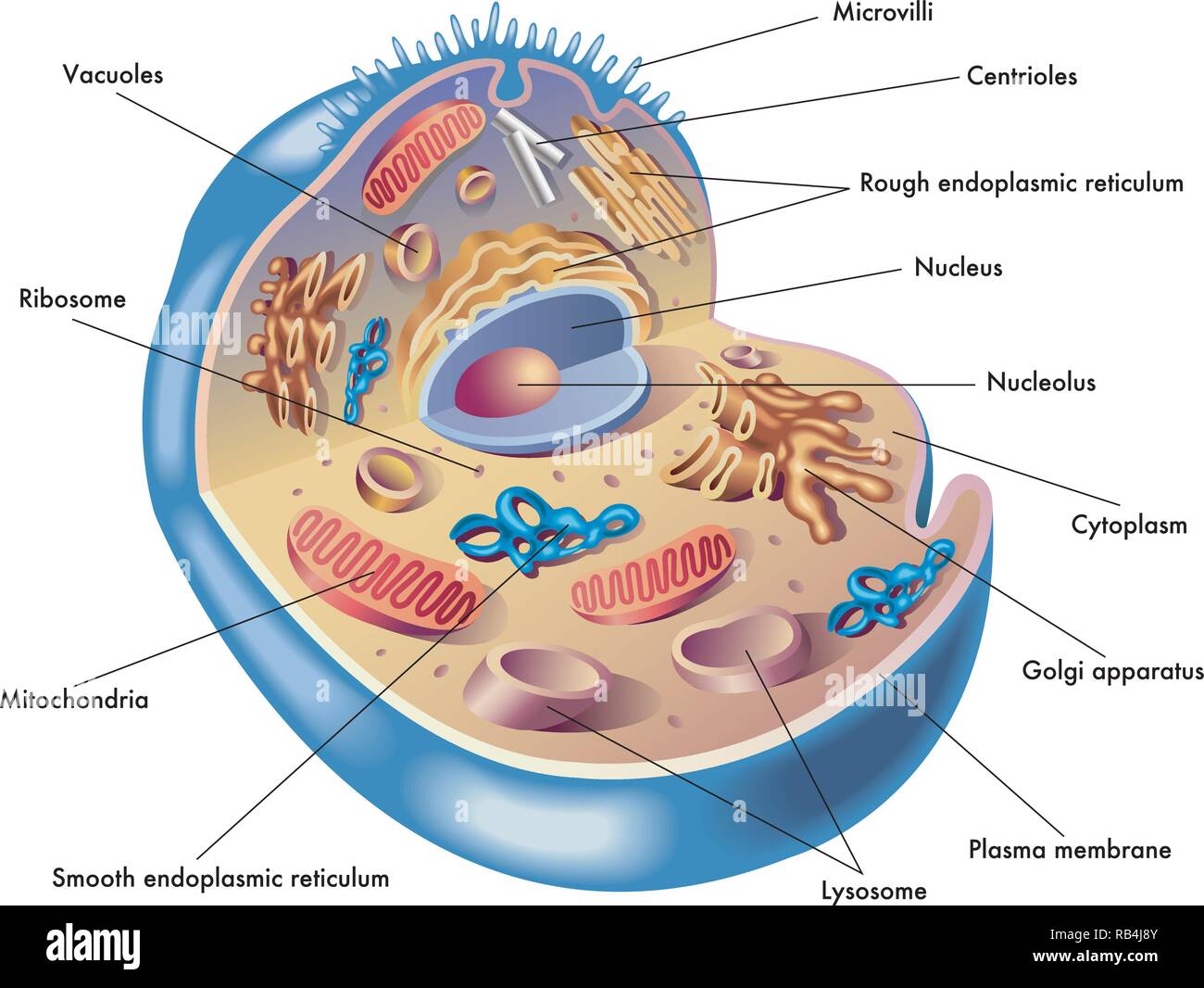
Medical illustration of elements of human cell Stock Vector Image & Art
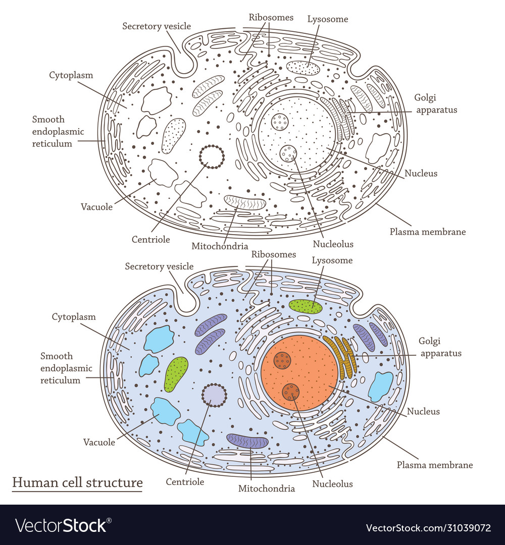
Human cell structure Royalty Free Vector Image
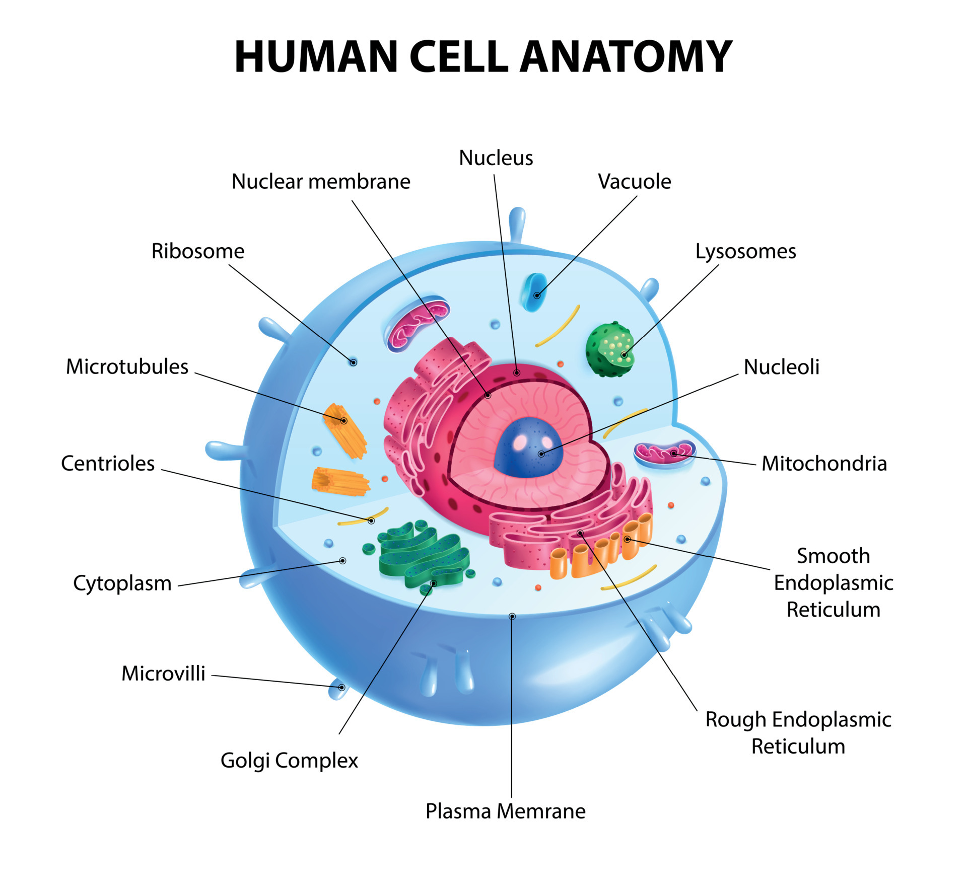
Human Cells
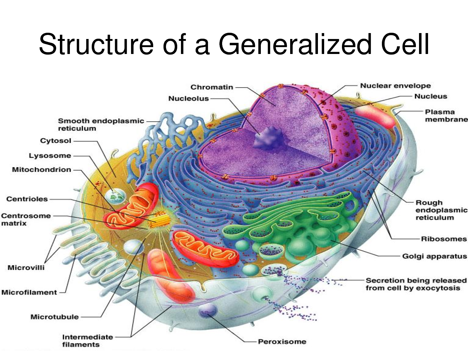
Human Cell Diagrams Images & Pictures Becuo

Human Cell Diagram, Parts, Pictures, Structure and Functions
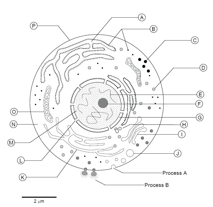
Human Cell Drawing at Explore collection of Human

Structure Of Human Cell With Labels Images & Pictures Becuo
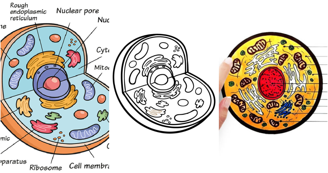
20 Easy Cell Drawing Ideas How to Draw a Cell

69,023 Human Cell Structure Images, Stock Photos & Vectors Shutterstock

How to Draw Human Cell Step by Step YouTube
Web Visual Guide To Human Cells.
The Cell Membrane Is The Outer Coating Of The Cell And Contains The Cytoplasm, Substances Within It And The Organelle.
Web Browse 5,800+ Human Cell Diagram Stock Photos And Images Available, Or Search For Cells To Find More Great Stock Photos And Pictures.
Scribble Around Inside The Cell With Wavy Lines Are Ovals.
Related Post: