Embryology Drawing
Embryology Drawing - This process will both help with repetition (see #3) and reinforce your understanding of the material. List and describe four embryonic membranes. Web embryo images normal and abnormal mammalian development is a tutorial that uses scanning electron micrographs (sems) as the primary resource to teach mammalian embryology. Distinguish the stages of embryonic development that occur before implantation. Stewart/spl) some of the most iconic images in biology hold a dark secret. Web we propose that the development of embryology resources to support visualisation can initiate a dialogue with the students of embryology, with the aim of enhancing teaching and learning practices. 3d models, histology sections and gene expression data in human embryonic and fetal development. Web embryo drawing is the illustration of embryos in their developmental sequence. Leonardo is considered to be the very first in history to correctly depict the human fetus in its proper position within the womb. In plants and animals, an embryo develops from a zygote, the single cell that results when an egg and sperm fuse during fertilization. A new book tells, for the first time in full, the extraordinary story of drawings of embryos initially published in 1868. Leonardo is considered to be the very first in history to correctly depict the human fetus in its proper position within the womb. This lesson concentrates on two aspects of using drawings to illustrate and record observations: In my. In plants and animals, an embryo develops from a zygote, the single cell that results when an egg and sperm fuse during fertilization. This period is also considered the organogenic period, when most organs within the embryo have begun to form. 3d models, histology sections and gene expression data in human embryonic and fetal development. This process will both help. Web a diagram showing real vertebrate embryos by michael richardson and colleagues (used in figure 4.2 of explore evolution) suggest that haeckel took considerable license in portraying the earlier embryos in the series, particularly in the top row. In plants and animals, an embryo develops from a zygote, the single cell that results when an egg and sperm fuse during. He was also the first to expertly draw the uterine artery and the vascular system of the cervix and vagina. A new book tells, for the first time in full, the extraordinary story of drawings of embryos initially published in 1868. Leonardo is considered to be the very first in history to correctly depict the human fetus in its proper. Web we propose that the development of embryology resources to support visualisation can initiate a dialogue with the students of embryology, with the aim of enhancing teaching and learning practices. Embryo images normal and abnormal mammalian development is a tutorial that uses scanning electron micrographs (sems) as the primary resource to teach mammalian embryology. Web leonardo believed that the fetus. This process will both help with repetition (see #3) and reinforce your understanding of the material. He was also the first to expertly draw the uterine artery and the vascular system of the cervix and vagina. Web a diagram showing real vertebrate embryos by michael richardson and colleagues (used in figure 4.2 of explore evolution) suggest that haeckel took considerable. Web a diagram showing real vertebrate embryos by michael richardson and colleagues (used in figure 4.2 of explore evolution) suggest that haeckel took considerable license in portraying the earlier embryos in the series, particularly in the top row. In my undergraduate cell biology class, we used modeling clay to build out cellular processes like dna transcription. Describe the process of. Web you can also make your own visual depictions of embryological processes by drawing them. Web wells implies that textbooks misrepresent u000bthe study of developmental programs as evidence for evolution by accusing them of using haeckel's inaccurate drawings,u000bin effect accusing textbooks that show any embryos of mindlessly repeating haeckel. Bulleted notes & final drawings. Leonardo is considered to be the. Web embryo drawing is the illustration of embryos in their developmental sequence. Distinguish the stages of embryonic development that occur before implantation. He was also the first to expertly draw the uterine artery and the vascular system of the cervix and vagina. Web you can also make your own visual depictions of embryological processes by drawing them. A new book. He was also the first to expertly draw the uterine artery and the vascular system of the cervix and vagina. Minimalist or cartoon drawings to just illustrate the points being made without distracting information; The images that would not go away. Embryo images normal and abnormal mammalian development is a tutorial that uses scanning electron micrographs (sems) as the primary. These early papers often included drawings of histological sections from specific embryos, providing a prelude to later photomicrographs. In my undergraduate cell biology class, we used modeling clay to build out cellular processes like dna transcription. Distinguish the stages of embryonic development that occur before implantation. List and describe four embryonic membranes. Web we propose that the development of embryology resources to support visualisation can initiate a dialogue with the students of embryology, with the aim of enhancing teaching and learning practices. In plants and animals, an embryo develops from a zygote, the single cell that results when an egg and sperm fuse during fertilization. In plants and animals, an embryo develops from a zygote, the single cell that results when an egg and sperm fuse during fertilization. Describe how the placenta is formed and identify its functions. This period is also considered the organogenic period, when most organs within the embryo have begun to form. 3d models, histology sections and gene expression data in human embryonic and fetal development. There are links to more detailed descriptions which can be viewed in a week by week format. The images that would not go away. Web embryo drawing is the illustration of embryos in their developmental sequence. Bulleted notes & final drawings. Web wells implies that textbooks misrepresent u000bthe study of developmental programs as evidence for evolution by accusing them of using haeckel's inaccurate drawings,u000bin effect accusing textbooks that show any embryos of mindlessly repeating haeckel. Web in one of his most famous drawings, leonardo depicts a human fetus lying inside a dissected uterus.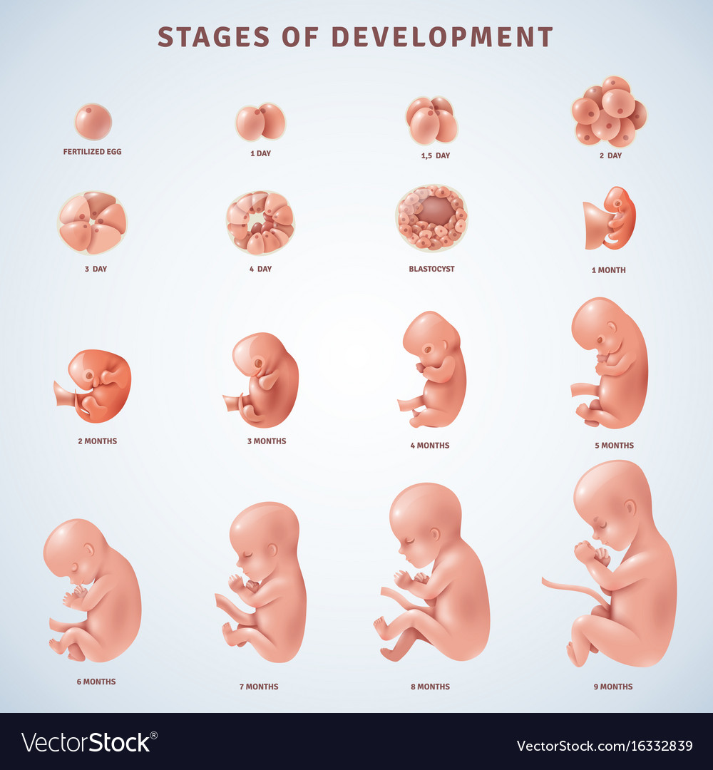
Stages human embryonic development Royalty Free Vector Image
Stages in human embryonic development 455531 Vector Art at Vecteezy

Paper Description of a 4 mm human embryo (1906) Embryology
Embryology Evolution
Human embryonic development in human infographic 6158571 Vector Art at
Human Embryo Evolution, Flat Vector Illustration. Fetal Development

Human embryo Royalty Free Vector Image VectorStock

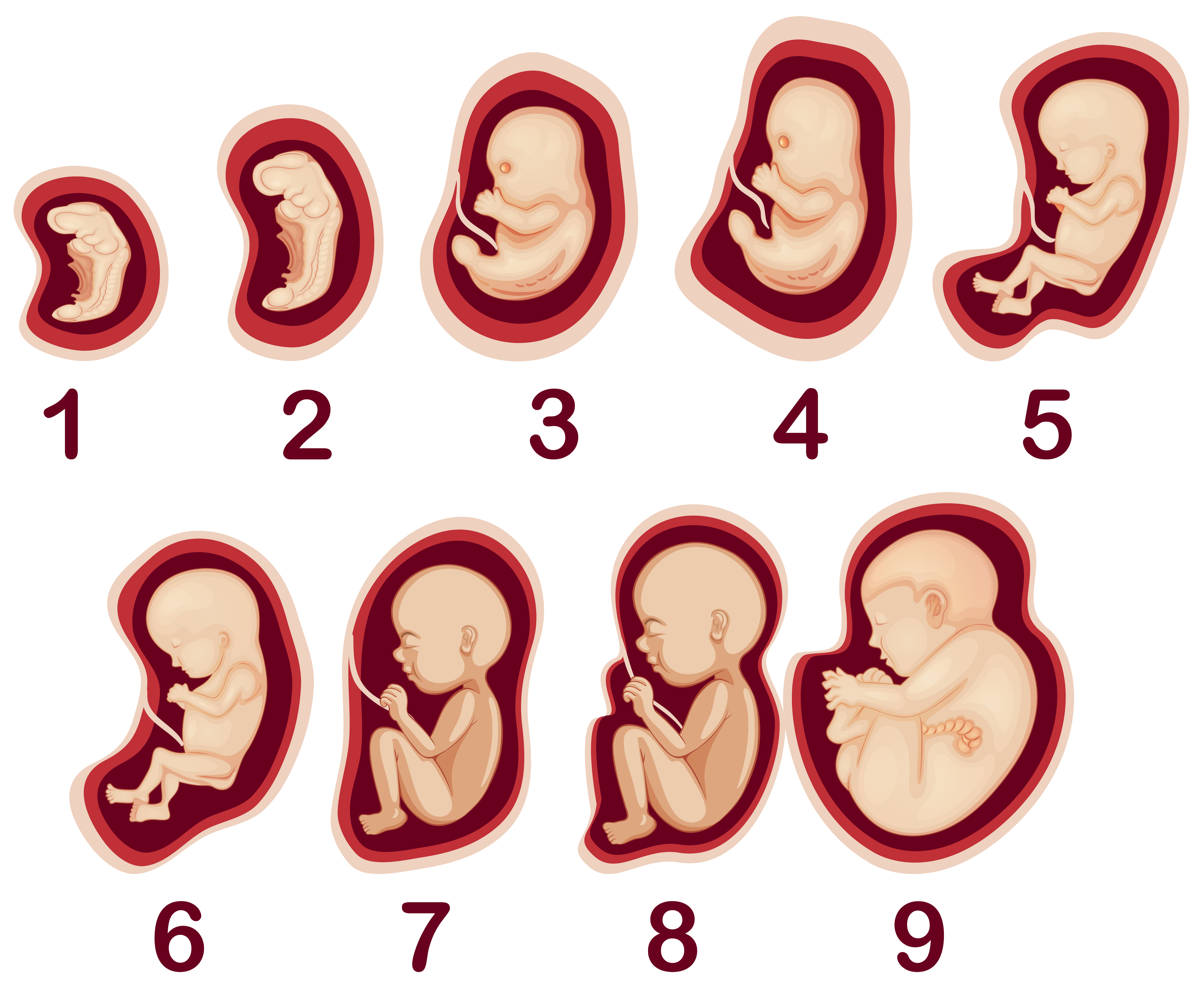
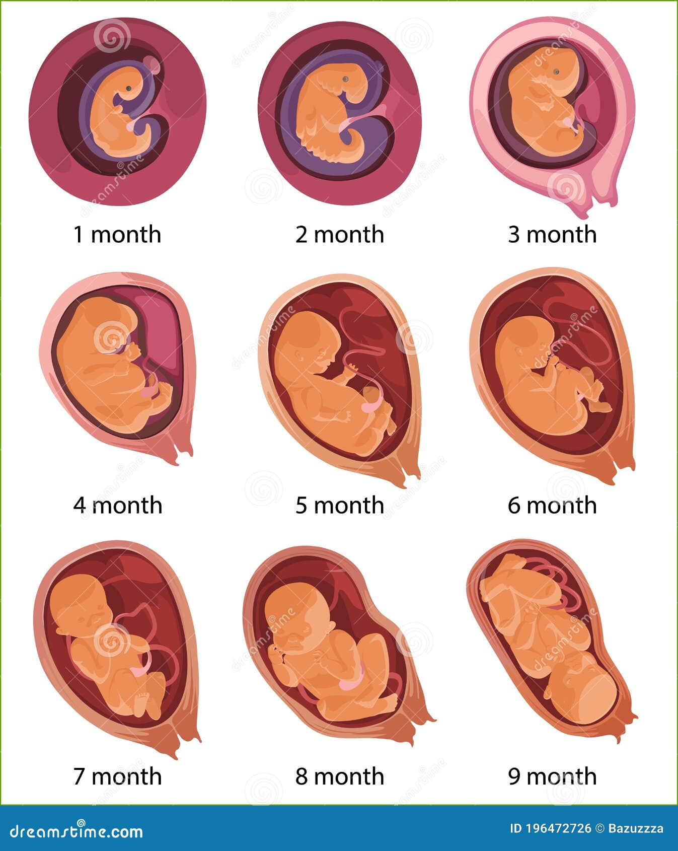
Embryo Development A Development process of Fetus Week by Week
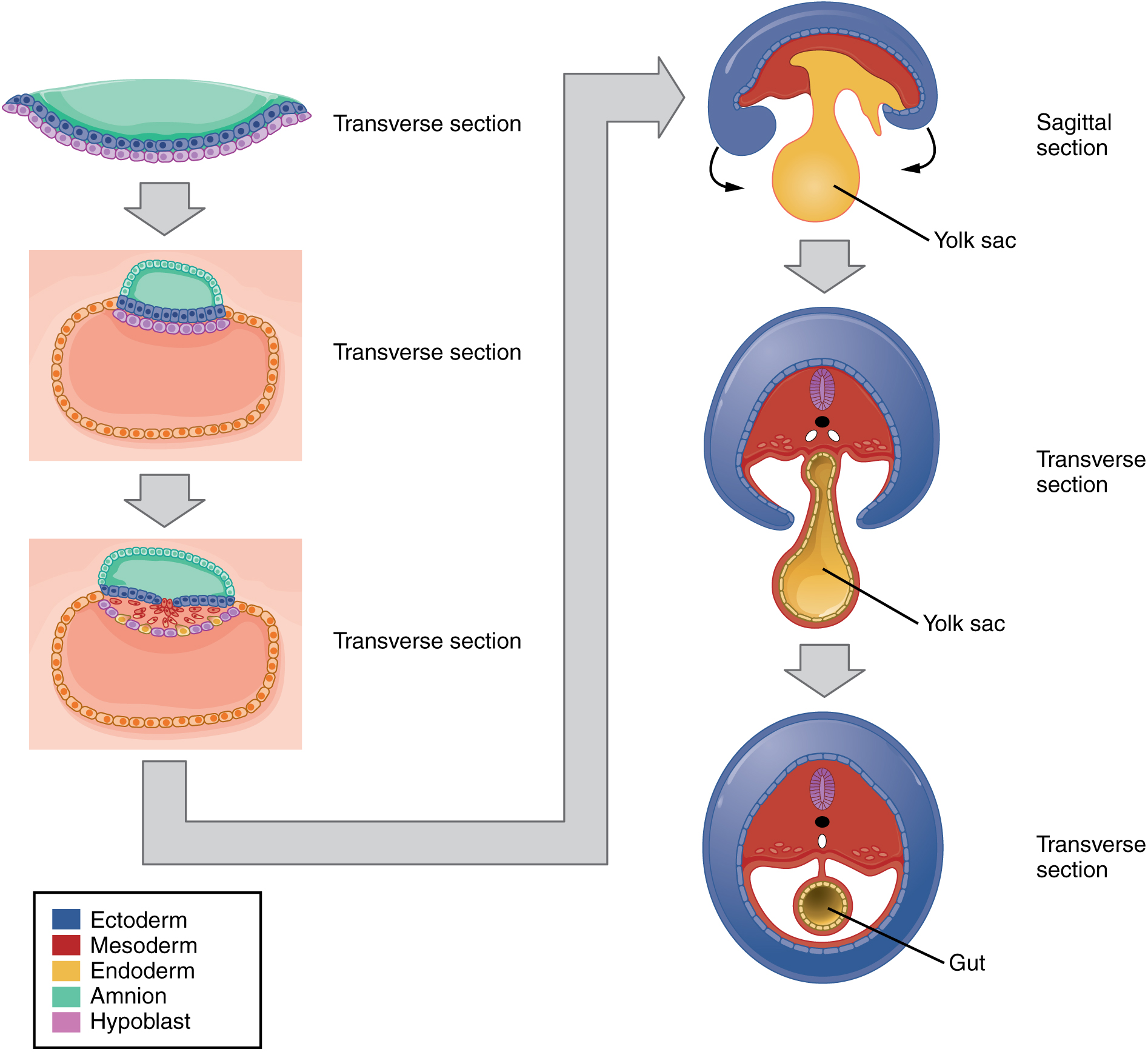
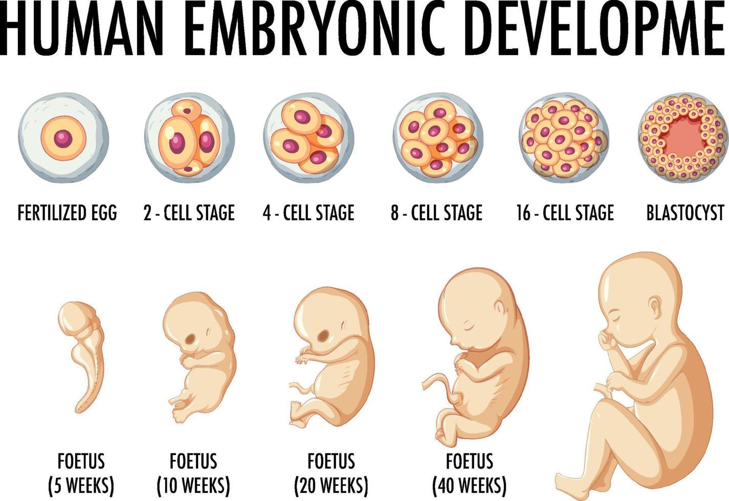
Embryonic Development · Anatomy and Physiology
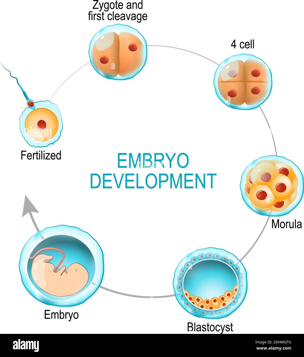
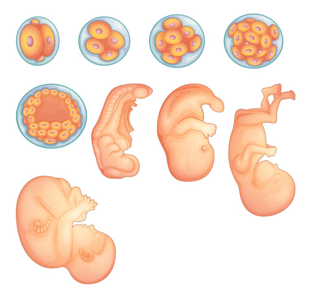
embryo development. from fertilization to zygote, morula and Blastocyst
Web You Can Also Make Your Own Visual Depictions Of Embryological Processes By Drawing Them.
By The End Of This Section, You Will Be Able To:
Stewart/Spl) Some Of The Most Iconic Images In Biology Hold A Dark Secret.
Leonardo Is Considered To Be The First To Have Quantitatively Measured The Fetus.
Related Post: