Drawing Of Esophagus
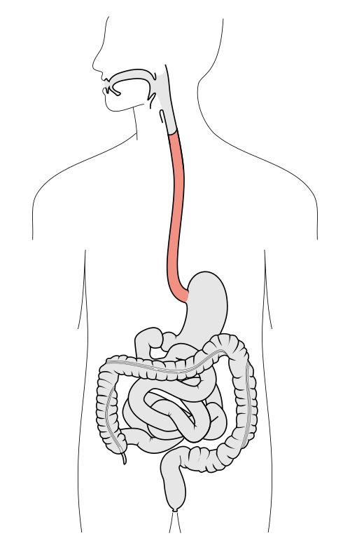
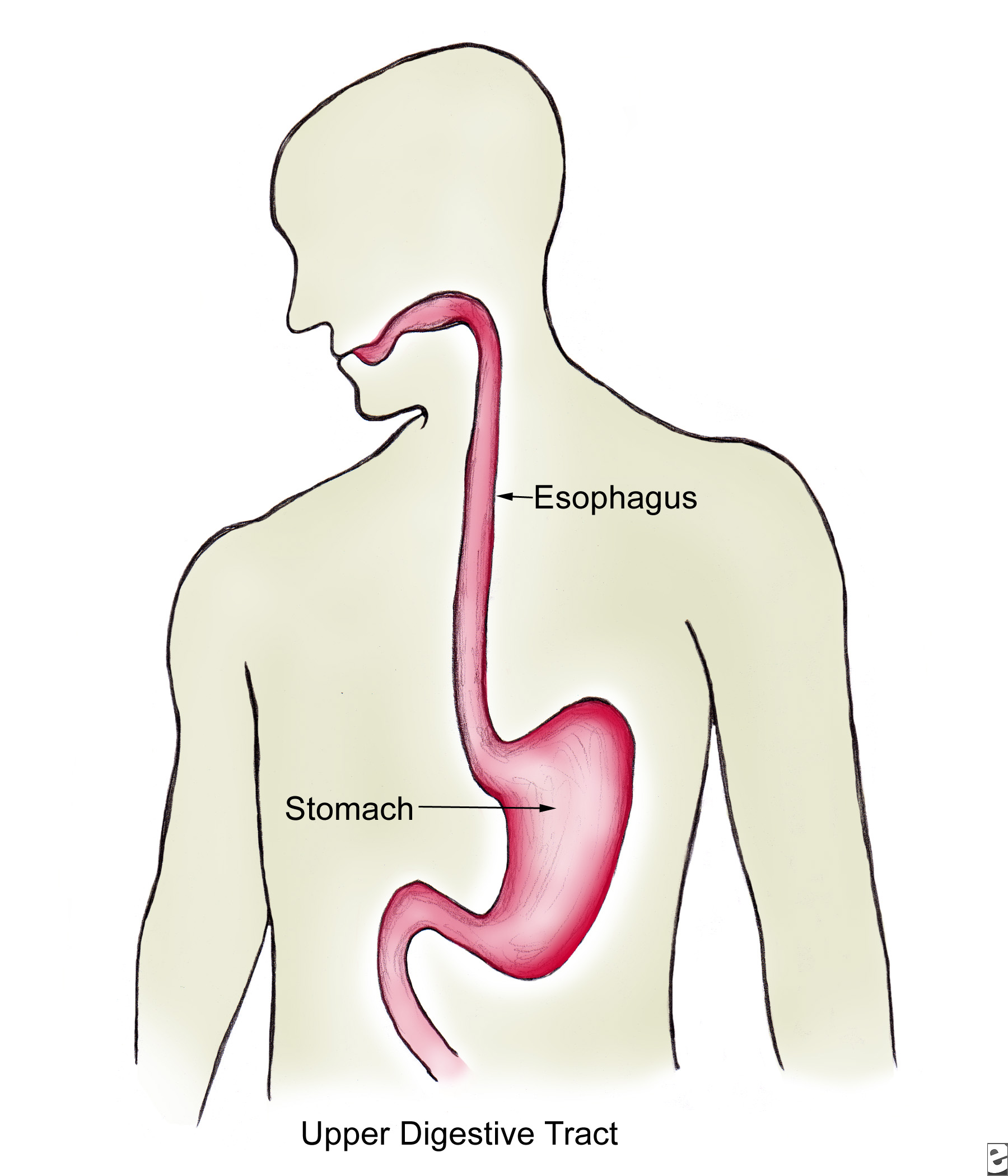
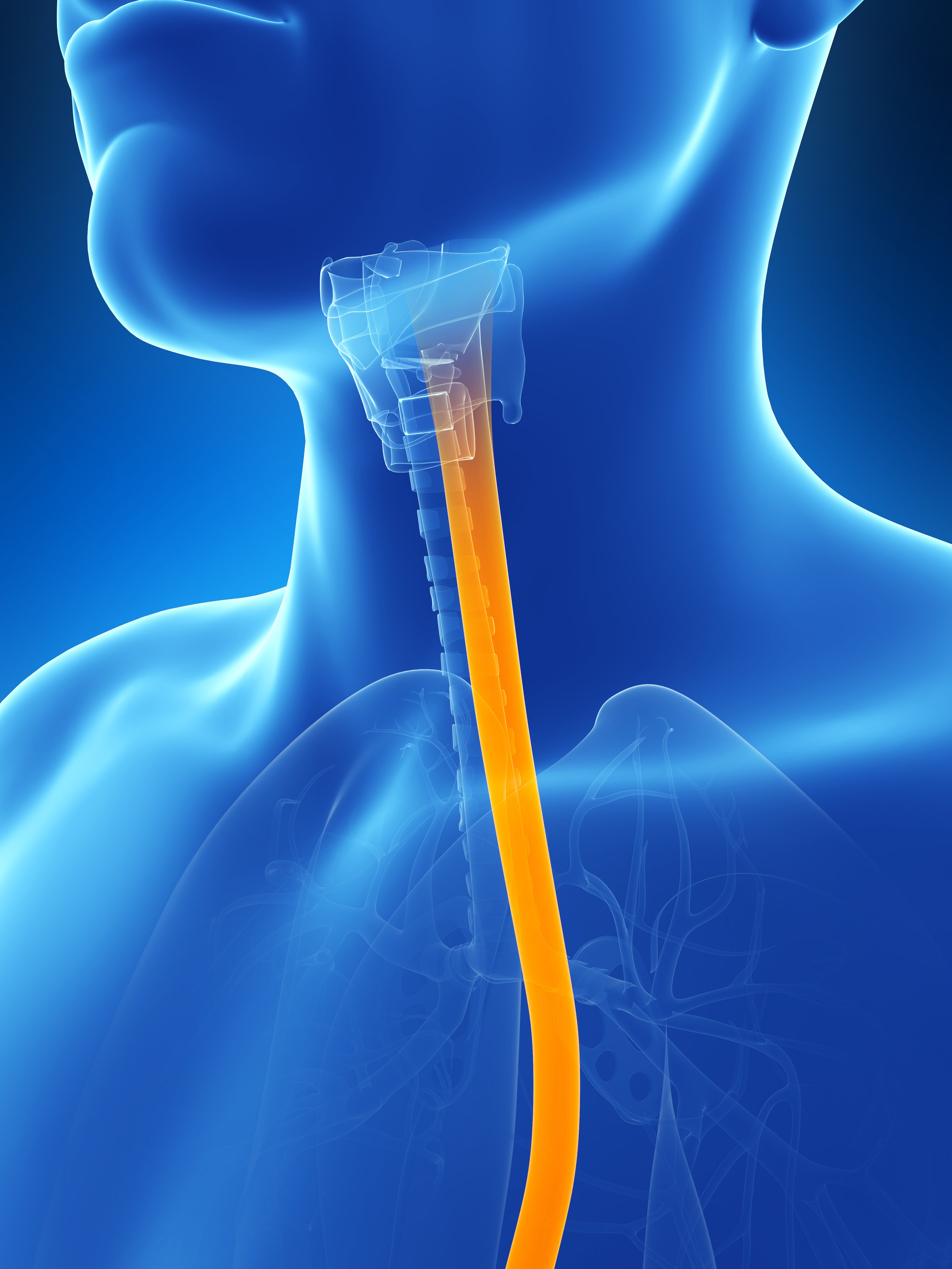
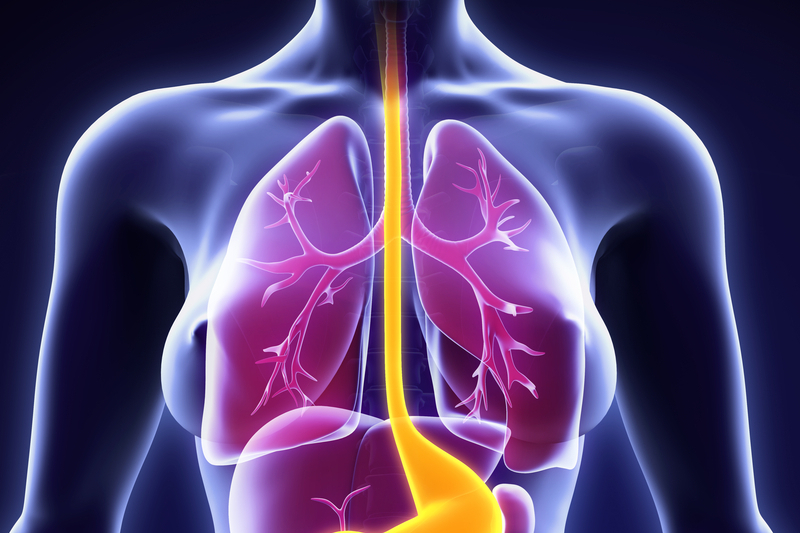
Drawing Of Esophagus - Long, extending from the pharynx to the stomach. It should be fairly narrow, about 1/5 the width of your model's neck. Web drawing of the digestive system with the mouth; The esophagus lies posterior to the trachea and the heart and passes through the mediastinum and the hiatus, an opening in the diaphragm, in its descent from the thoracic to the abdominal cavity. Sometimes it progresses to a more serious condition called. Browse 71 esophagus drawing photos and images available, or start a new search to explore more photos and images. So i'll draw three parts of the esophagus here. The oesophagus or gullet is a muscular canal, about 23 to 25 cm. And so that's 1/3, i'll say the top 1/3 of the esophagus. Web the esophagus is a long, thin, and muscular tube that connects the pharynx (throat) to the stomach. Web an intimate gathering at lulu at the hammer museum on april 15 hailed a milestone for the ucla robert g. Web drawing of the digestive system with the mouth; Browse 71 esophagus drawing photos and images available, or start a new search to explore more photos and images. When we swallow food or liquids the epiglottis falls back and. Web several other structures border the esophagus throughout its journey, so here’s an esophageal diagram that provides you with an overview of all of them! Web draw the esophagus. Web acid reflux, which affects nearly a third of u.s. Colon, also called the large intestine; The esophagus lies posterior to the trachea and the heart and passes through the mediastinum. Adults weekly, is the backward flow of stomach acid into the esophagus. Web choose from drawing of esophagus stock illustrations from istock. Find a primary care provider. Web choose from 275 drawing of a esophagus stock illustrations from istock. Healthy stomach inside man body vector. Sometimes it progresses to a more serious condition called. The cheeks, tongue, and palate frame the mouth, which is also called the oral cavity (or buccal cavity). The esophagus lies posterior to the trachea and the heart and passes through the mediastinum and the hiatus, an opening in the diaphragm, in its descent from the thoracic to the abdominal cavity.. Web several other structures border the esophagus throughout its journey, so here’s an esophageal diagram that provides you with an overview of all of them! It should be fairly narrow, about 1/5 the width of your model's neck. Extends from the pharynx to the stomach, anterior to the vertebral bodies of the vertebral column. Web choose from drawing of esophagus. The esophagus is the hollow, muscular tube that passes food and liquid from your throat to your stomach. Web acid reflux, which affects nearly a third of u.s. Web draw the esophagus. Web browse 236 drawing of esophagus stock photos and images available, or start a new search to explore more stock photos and images. Find a primary care provider. It's split up into thirds. Web an intimate gathering at lulu at the hammer museum on april 15 hailed a milestone for the ucla robert g. Find a primary care provider. Healthy stomach inside man body vector. So the first part that we have is actually just skeletal muscle. The food moves from the mouth into the esophagus, which carries it down into the stomach. Browse 71 esophagus drawing photos and images available, or start a new search to explore more photos and images. Web drawing of the gi tract, with the esophagus, stomach, small intestine, duodenum, jejunum, ileum, large intestine, cecum, colon, rectum, and anus labeled. Web choose. The cheeks, tongue, and palate frame the mouth, which is also called the oral cavity (or buccal cavity). Web drawing of the digestive system with the mouth; Web the esophagus is a hollow tube that begins at the back of the mouth at around the sixth cervical vertebrae. Lamina muscularis layer of esophagus. Web several other structures border the esophagus. The cheeks, tongue, and palate frame the mouth, which is also called the oral cavity (or buccal cavity). Colon, also called the large intestine; Long, extending from the pharynx to the stomach. Web the esophagus is a long, thin, and muscular tube that connects the pharynx (throat) to the stomach. Its main job is to deliver food, liquids, and saliva. The esophagus is the muscular tube that connects the back of the throat (or pharynx) with the stomach. Web browse 236 drawing of esophagus stock photos and images available, or start a new search to explore more stock photos and images. Extends from the pharynx to the stomach, anterior to the vertebral bodies of the vertebral column. Find a primary care provider. Lamina propria of mucosa layers of esophagus. Web choose from 275 drawing of a esophagus stock illustrations from istock. The food moves from the mouth into the esophagus, which carries it down into the stomach. Lymhatic nodules (not in all amimals) oin lamina propria layer. The cheeks, tongue, and palate frame the mouth, which is also called the oral cavity (or buccal cavity). And so that's 1/3, i'll say the top 1/3 of the esophagus. At the end of the mouth, draw a small tube that extends straight down into the center of your model’s torso. Choose from drawing of the esophagus stock illustrations from istock. So i'll draw three parts of the esophagus here. Lamina muscularis layer of esophagus. When we swallow food or liquids the epiglottis falls back and covers the larynx, sort of like a railroad switch, so food does not travel down into the bronchial passages of the lungs. Human digestive system woman anatomy diagram.
Esophagus Libre Pathology

The Mouth, Pharynx, and Esophagus Biology of Aging

The esophagus Structure of the esophagus

The Human Esophagus Functions and Anatomy and Problems

Esophagus / Endoscopy — High Plains Surgical Associates

Anatomy Of The Esophagus

Esophagus Facts, Functions & Diseases Live Science
![[DIAGRAM] Labeled Diagram Of The Esophagus](https://thumbor.kenhub.com/rpjXaMtj_rsixQh2Se_wOcXdwrI=/fit-in/800x1600/filters:watermark(/images/logo_url.png,-10,-10,0)/images/library/311/Esophagus.png)
[DIAGRAM] Labeled Diagram Of The Esophagus
:watermark(/images/watermark_5000_10percent.png,0,0,0):watermark(/images/logo_url.png,-10,-10,0):format(jpeg)/images/overview_image/292/Gspt830scLPX0uk5rXr0w_esophagus-in-situ_english.jpg)
Esophagus Anatomy, sphincters, arteries, veins, nerves Kenhub

E.3. Esophagus
Web In This Section, You Will Examine The Anatomy And Functions Of The Three Main Organs Of The Upper Alimentary Canal—The Mouth, Pharynx, And Esophagus—As Well As Three Associated Accessory Organs—The Tongue, Salivary Glands, And Teeth.
The Esophagus Lies Posterior To The Trachea And The Heart And Passes Through The Mediastinum And The Hiatus, An Opening In The Diaphragm, In Its Descent From The Thoracic To The Abdominal Cavity.
Many Regard The Esophagus As Merely A “Food Pipe” Through Which Food Traverses The Gap Between The Pharynx And The Stomach.
Web Draw The Esophagus.
Related Post: