Cranial Nerves Drawing
Cranial Nerves Drawing - Web the simplest way to draw the #cranial_nerves. Web the cranial nerves provide afferent and efferent innervation principally to the structures of the head and neck. #cranialnerveexam #cranialnerves #cranialnerve welcome back! Web diagrams of cranial nerves. This video is an overview of the. Central nerves are in your brain and spinal cord. Then, between the cerebral peduncles, draw the hypothalamus. Web draw the thalami, the major sensory integration center. The first two nerves (olfactory and optic) arise from the cerebrum, whereas the remaining ten emerge from the brainstem. Unlike spinal nerves, whose roots are neural fibers from the spinal grey matter, cranial nerves are composed of the neural processes associated with distinct brainstem nuclei and cortical structures. What are the cranial nerves? Then, between the cerebral peduncles, draw the hypothalamus. #cranialnerveexam #cranialnerves #cranialnerve welcome back! The first, the olfactory nerve, is responsible for smell. Cranial nerves are pairs of nerves that connect your brain to different parts of your head, neck, and trunk. What are the 12 cranial nerves? Olfactory bulb and tract (purple) optic nerve and chiasma (dark green) oculomotor (dark blue) trochlear (gray) trigeminal (pink) abducens (orange) Web the cranial nerves provide afferent and efferent innervation principally to the structures of the head and neck. Web schematic drawing of cranial nerve nuclei with vestibular complex shown. Web the human body has. Easy way to remember the 12 pairs of cranial nerves. Web diagrams of cranial nerves. Each nerve has a corresponding roman. These original anatomical drawings were produced digitally, working from medical imaging sources and 3d reconstructions using adobe illustrator. The first, the olfactory nerve, is responsible for smell. These signals help you smell, taste, hear and move your facial muscles. Olfactory nerve (cn i) optic nerve (cn ii) oculomotor nerve (cn iii) trochlear nerve (cn iv) trigeminal nerve (cn v) abducens nerve (cn vi) facial nerve (cn vii) vestibulocochlear nerve (cn viii) glossopharyngeal nerve (cn ix) vagus nerve (cn x) accessory nerve (cn xi) hypoglossal nerve (cn xii). Specify the pituitary stalk (in midline) and then the mammillary bodies. Web cranial nerves are the 12 nerves of the peripheral nervous system that emerge from the foramina and fissures of the cranium. A number of cranial nerves send electrical signals between your brain and different parts of your neck, head and torso. Web cranial nerve 12. The cranial nerves. The first two nerves (olfactory and optic) arise from the cerebrum, whereas the remaining ten emerge from the brainstem. They are a key part of your nervous system. Web functions and diagram. Web the human body has 12 pairs of cranial nerves that control motor and sensory functions of the head and neck. In this video i will go over. Web the cranial nerves provide afferent and efferent innervation principally to the structures of the head and neck. Color each part according to the keys. All cranial nerves originate from nuclei in the brain. Web the sheep brain below has many parts labeled and shows the cranial nerves. Easy way to remember the 12 pairs of cranial nerves. Web the human body has 12 pairs of cranial nerves that control motor and sensory functions of the head and neck. Web the cranial nerves are a set of 12 paired nerves that arise directly from the brain. Easy way to remember the 12 pairs of cranial nerves. This article provides a pictorial overview of the imaging of cranial nerves,. The optic nerve, cn ii, conveys visual information. Name the twelve cranial nerves and explain the functions associated with each. They are a key part of your nervous system. These original anatomical drawings were produced digitally, working from medical imaging sources and 3d reconstructions using adobe illustrator. The cranial nerves begin toward the back of your brain. Web the human body has 12 pairs of cranial nerves that control motor and sensory functions of the head and neck. Web the cranial nerves provide afferent and efferent innervation principally to the structures of the head and neck. Now, let's show the cranial nerves as they exit the brainstem. Web schematic drawing of cranial nerve nuclei with vestibular complex. Specify the pituitary stalk (in midline) and then the mammillary bodies. Web the human body has 12 pairs of cranial nerves that control motor and sensory functions of the head and neck. Web the cranial nerves provide afferent and efferent innervation principally to the structures of the head and neck. In this video i will go over cranial nerves i through twelve by drawing a picture to help you re. Web diagrams of cranial nerves. This section of the website will explain large and minute details of cranial nerves cross sectional anatomy. Then, between the cerebral peduncles, draw the hypothalamus. This video is an overview of the. Web the sheep brain below has many parts labeled and shows the cranial nerves. What are the 12 cranial nerves? Now, let's show the cranial nerves as they exit the brainstem. Web this mri cranial nerves cross sectional anatomy tool is absolutely free to use. They are a key part of your nervous system. Web here's a drawing of the brain looking up from the bottom, and all of these long stingy looking things coming out of the brain are cranial nerves. Web schematic drawing of cranial nerve nuclei with vestibular complex shown. Olfactory nerve (cn i) optic nerve (cn ii) oculomotor nerve (cn iii) trochlear nerve (cn iv) trigeminal nerve (cn v) abducens nerve (cn vi) facial nerve (cn vii) vestibulocochlear nerve (cn viii) glossopharyngeal nerve (cn ix) vagus nerve (cn x) accessory nerve (cn xi) hypoglossal nerve (cn xii) blood vessels &.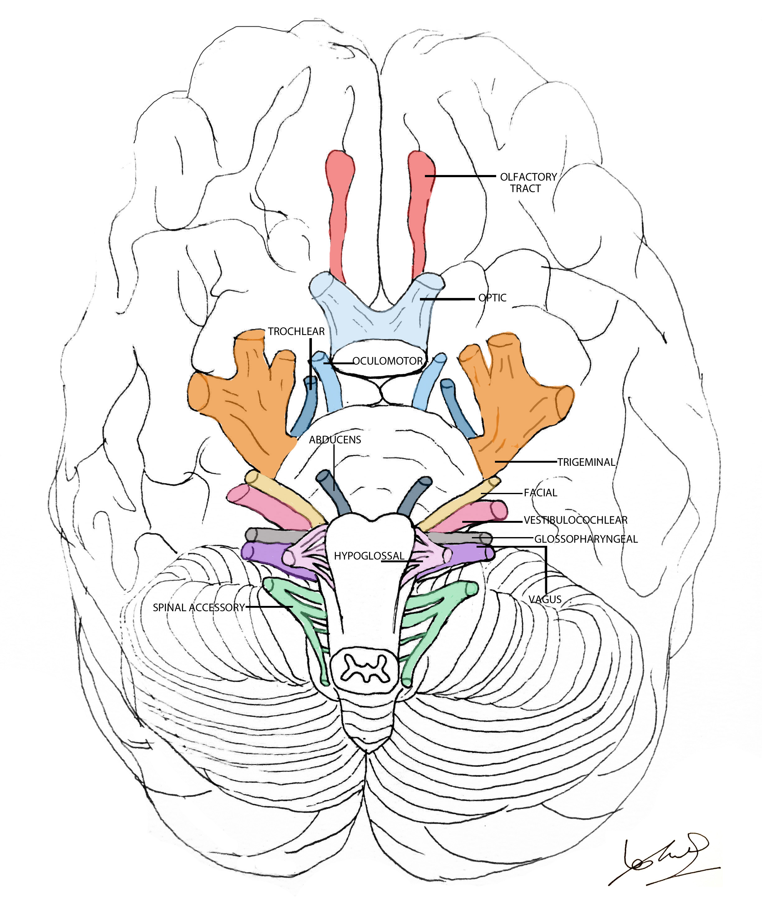
Cranial nerves explained Geeky Medics
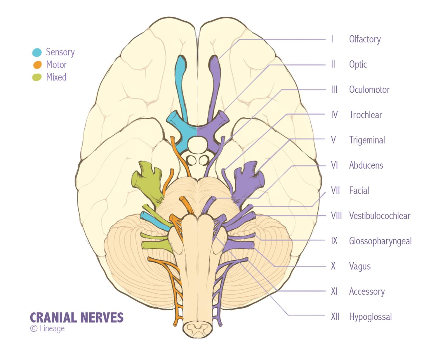
Cranial Nerves Neurology Medbullets Step 1
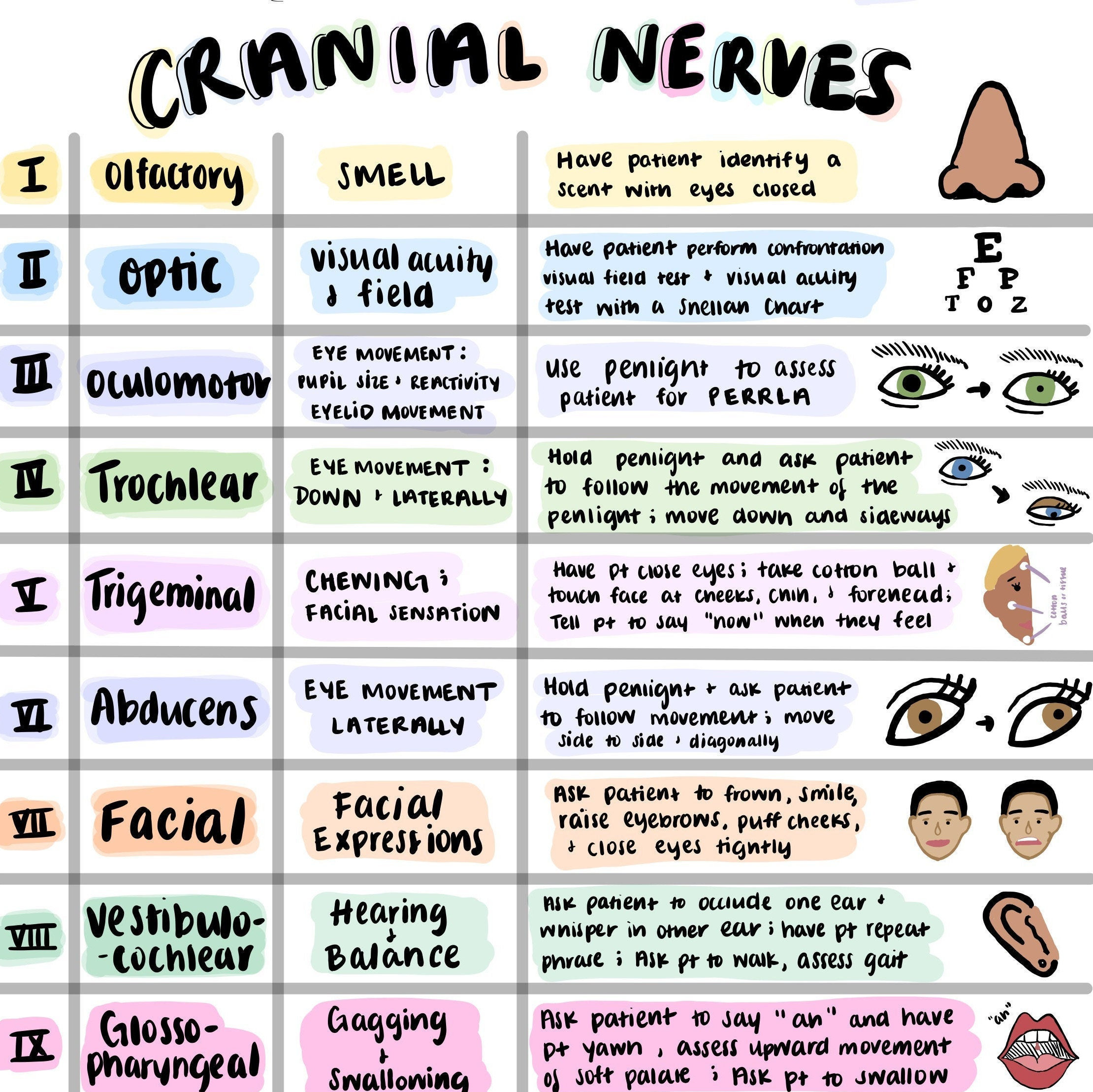
Cranial Nerves Sheet Colorful Hand Drawn Pictures for Etsy Ireland

Illustration of Cranial Nerves Stock Image F031/5295 Science

Cranial Nerves, Illustration Stock Image C030/5939 Science Photo

Cranial Nerves Drawing at GetDrawings Free download
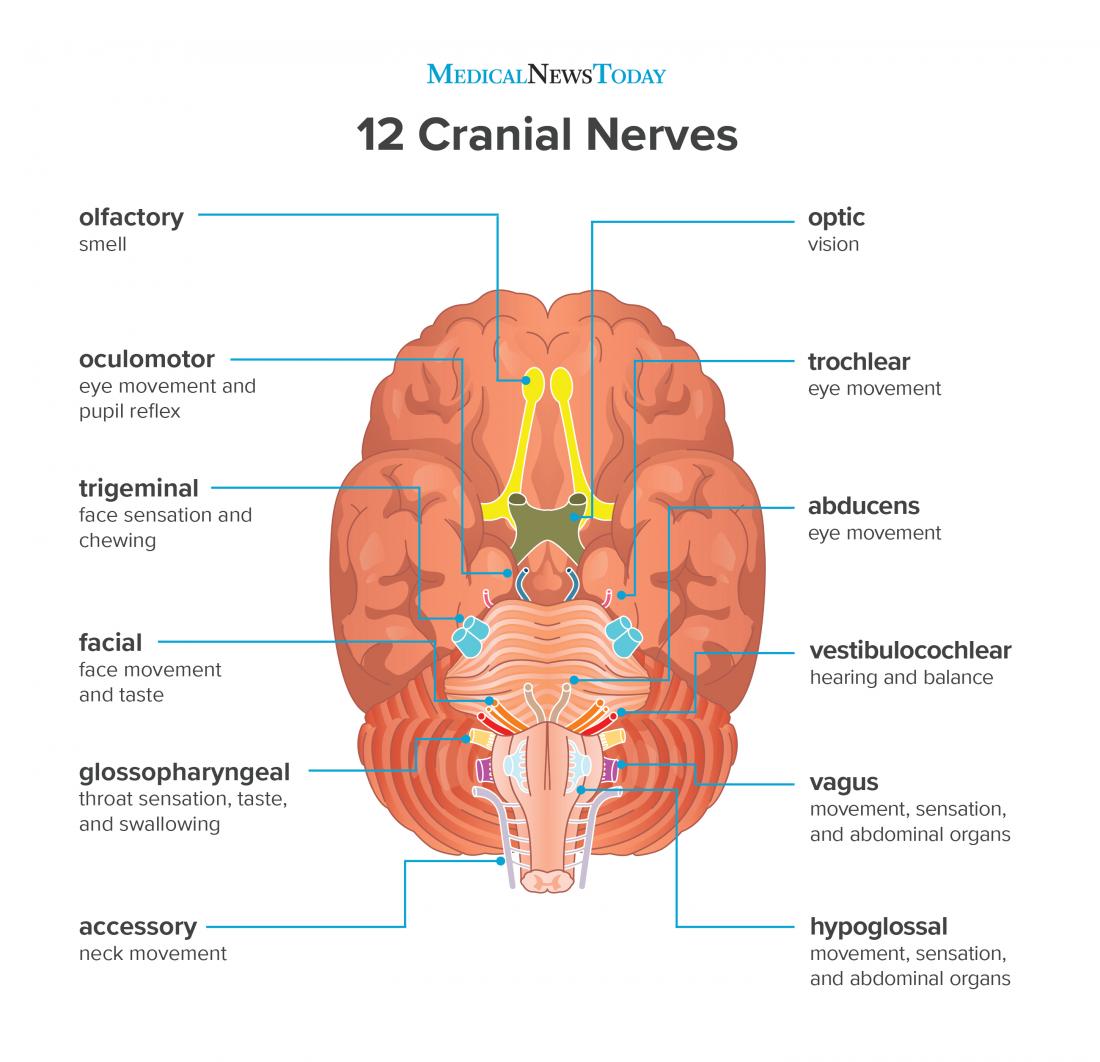
What are the 12 cranial nerves? Functions and diagram
:max_bytes(150000):strip_icc()/GettyImages-141483691-4cc225237a5945f8ab949d936f52c48e.jpg)
Cranial Nerves Anatomy, Function, and Treatment
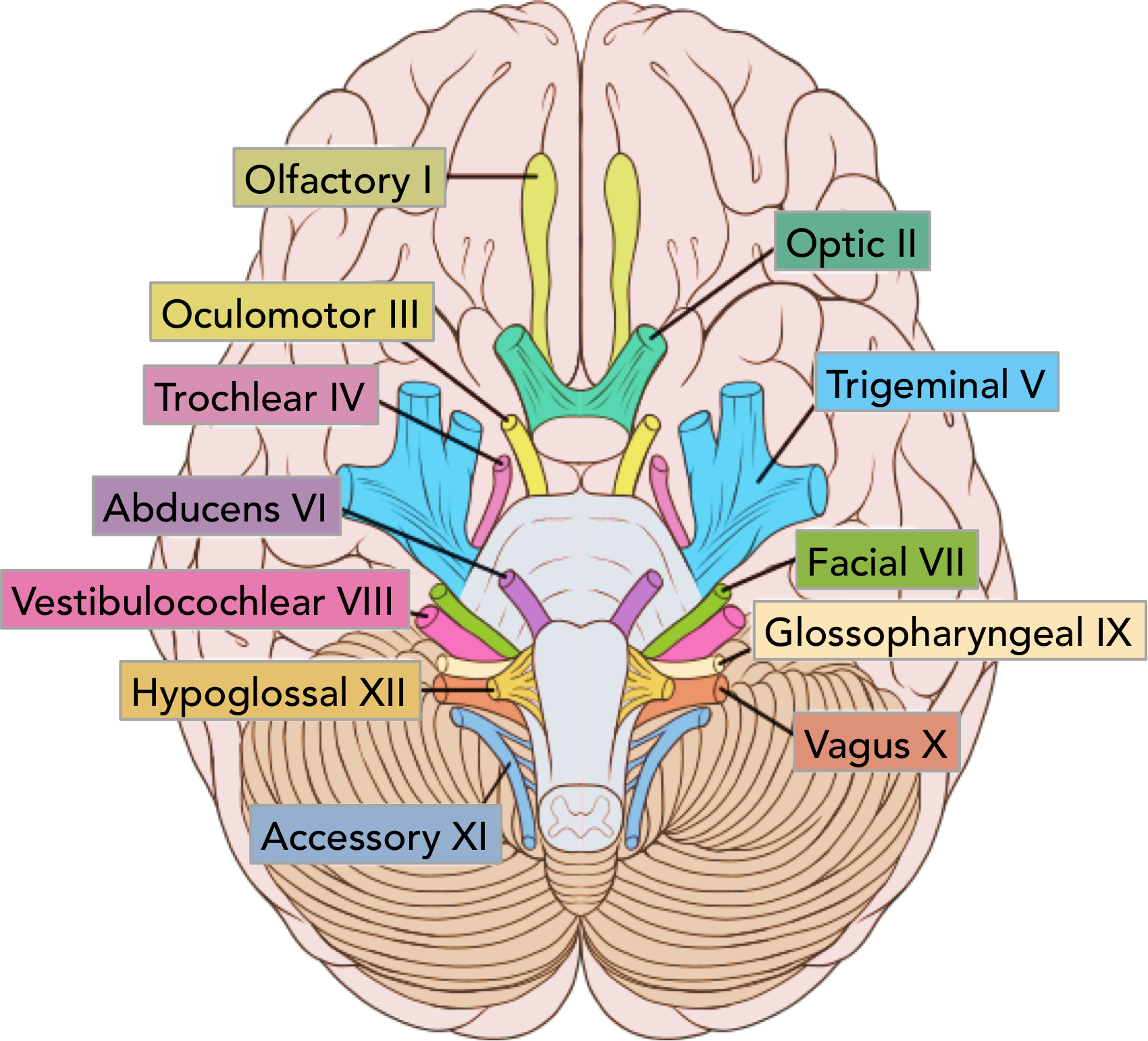
Summary of the Cranial Nerves TeachMeAnatomy
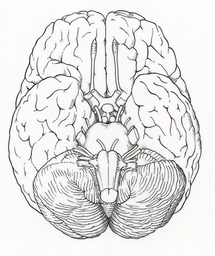
Cranial Nerves Drawing at GetDrawings Free download
The Optic Nerve, Cn Ii, Conveys Visual Information.
Describe The Sensory And Motor Components Of Spinal Nerves And The Plexuses That They Pass Through.
Web Cranial Nerve 12.
Next, The Oculomotor Nerve Moves The Eye Along With Cn Iv, The Trochlear Nerve And Cn Vi, The Abducens Nerve.
Related Post: