Bile Duct Drawing
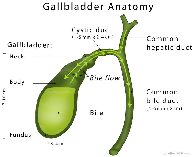
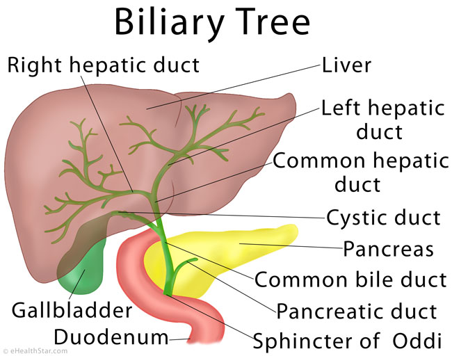
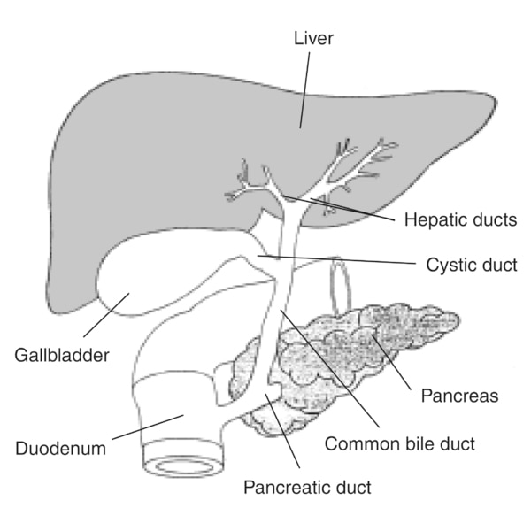
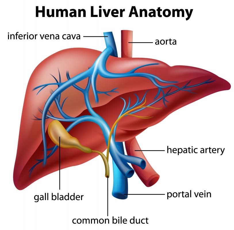
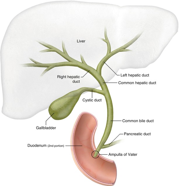
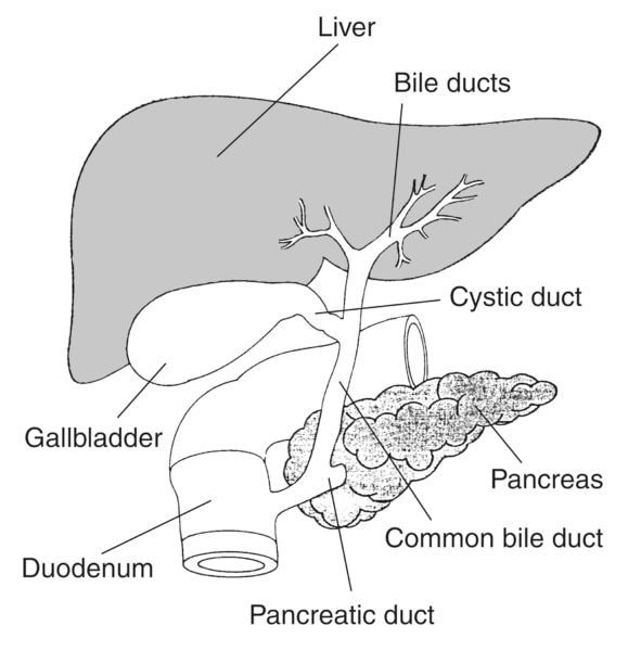
Bile Duct Drawing - During digestion, bile is released by the gallbladder into the cystic duct. Drawing of the biliary system with the liver, biliary tree (bile ducts), common bile duct, gallbladder, pancreas, duodenal papilla, main pancreatic duct, and duodenum. Drawing of the biliary system, with the liver, gallbladder, duodenum, pancreatic duct, common bile duct, pancreas, cystic duct, and hepatic ducts labeled. The bile will look similar to fluid in density. Drawing shows the liver and the intrahepatic bile ducts, which include the right and left hepatic ducts. 354 kb | 1693 x 1069. Web anatomy of the intrahepatic bile ducts; Web the common bile duct is formed by the junction of the cystic and hepatic ducts; Their purpose is to carry bile between these organs. Web canalicular bile flow is stimulated by bile acids, while ductular/ductal bile flow is stimulated by secretin. Neurovascular supply and lymphatic drainage of the gallbladder. Web anatomy of the intrahepatic bile ducts; It then runs in a groove near the. 742 kb | 2025 x 2100. Drawing of the biliary system, with the liver, gallbladder, duodenum, pancreatic duct, common bile duct, pancreas, cystic duct, and hepatic ducts labeled. This is a small, hollow tube that functions to transport bile. Web anatomy of the intrahepatic bile ducts; Web the transportation of bile follows this sequence: A bile duct drain, also called biliary drainage, can. A normal gallbladder will have a thin, barely perceptible wall, and will not appear dilated. Web a bile duct drain is a procedure that involves opening up obstructions or treating holes in the biliary system. What is a bile duct drain? Drawing of the biliary system with the liver, biliary tree (bile ducts), common bile duct, gallbladder, pancreas, duodenal papilla, main pancreatic duct, and duodenum labeled. In hepatic surgery, a preoperative understanding of bile duct. Web figure 21.7.4 is a drawing showing the anterior view of the gallbladder, part of the liver superior to the gallbladder, and the duct connecting the liver to gallbladder, eventually becoming the common bile duct. Drawing shows the extrahepatic bile ducts, including the common hepatic duct (perihilar region) and the common bile duct (distal region). When the liver cells secrete. Web a bile duct drain is a procedure that involves opening up obstructions or treating holes in the biliary system. Web the biliary tree is a complex network of conduits that begins with the canals of hering and progressively merges into a system of interlobular, septal, and major ducts which then coalesce to form the extrahepatic bile ducts, which finally. A normal gallbladder will have a thin, barely perceptible wall, and will not appear dilated. The biliary ducts combine to form the left and right hepatic ducts, which in turn combine to form the common hepatic duct. There is significant variation in the biliary tree with the classical description below thought to be present in ~60% of the population 2.. Bile canaliculi unite to form segmental bile ducts which drain each liver segment. Web what is a bile duct? Some patients have calcified gallstones that are visible on ct and look like small bright dots. This is a small, hollow tube that functions to transport bile. These ducts ultimately drain into the common hepatic duct. Their purpose is to carry bile between these organs. This is a small, hollow tube that functions to transport bile. There is significant variation in the biliary tree with the classical description below thought to be present in ~60% of the population 2. Web canalicular bile flow is stimulated by bile acids, while ductular/ductal bile flow is stimulated by secretin.. Also shown is the common hepatic duct, gallbladder, cystic duct, common bile duct, pancreas, ampulla of. 354 kb | 1693 x 1069. A normal gallbladder will have a thin, barely perceptible wall, and will not appear dilated. Web the biliary tree is a complex network of conduits that begins with the canals of hering and progressively merges into a system. There is significant variation in the biliary tree with the classical description below thought to be present in ~60% of the population 2. This is a small, hollow tube that functions to transport bile. Drawing of the biliary system with the liver, biliary tree (bile ducts), common bile duct, gallbladder, pancreas, duodenal papilla, main pancreatic duct, and duodenum. Inset of. Left and right hepatic ducts, 4. Web figure 21.7.4 is a drawing showing the anterior view of the gallbladder, part of the liver superior to the gallbladder, and the duct connecting the liver to gallbladder, eventually becoming the common bile duct. This is a small, hollow tube that functions to transport bile. Water movement in the bile ducts or ductules occurs through aquaporin 1, 41 which does not transport mannitol. Drawing of the biliary system with the liver, biliary tree (bile ducts), common bile duct, gallbladder, pancreas, duodenal papilla, main pancreatic duct, and duodenum labeled. 192 kb | 780 x 660. 354 kb | 1693 x 1069. Web 135 kb | 600 x 951. Drawing of the biliary system with the liver, biliary tree (bile ducts), common bile duct, gallbladder, pancreas, duodenal papilla, main pancreatic duct, and duodenum. Neurovascular supply and lymphatic drainage of the gallbladder. Web bile leaves the liver via biliary ducts continuously and is stored in the gall bladder until needed. These ducts ultimately drain into the common hepatic duct. Web anatomy of the intrahepatic bile ducts; Drawing of the biliary system, with the liver, gallbladder, duodenum, pancreatic duct, common bile duct, pancreas, cystic duct, and hepatic ducts labeled. It is about 7.5 cm. In hepatic surgery, a preoperative understanding of bile duct variation will help avoid possible complications and help achieve the most effective relief of chd obstruction.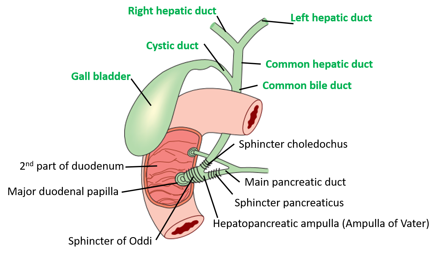
Extrahepatic Biliary Apparatus Anatomy QA

Biliary Tree Anatomy Bile duct, Human digestive system, Gallbladder
Bile Production, Secretion, Flow, Storage, Composition, pH, Function

biliary tract Liver anatomy, Diagnostic medical sonography, Medical
Liver Anatomy, Location and Function eHealthStar

(Color online) Anatomy of the biliary system. Download Scientific Diagram
Anatomy of the Biliary System with Labels Media Asset NIDDK
What is a Bile Duct? (with pictures)
Bile Duct Diagram
Biliary system with the liver, gallbladder, pancreas, duodenum, bile
742 Kb | 2025 X 2100.
Gallstones Are The Most Common Cause Of Bile Duct Obstructions.
Anatomy Of The Extrahepatic Bile Ducts;
Web The Common Bile Duct Is Formed By The Junction Of The Cystic And Hepatic Ducts;
Related Post: