Anatomical Drawing Of The Knee
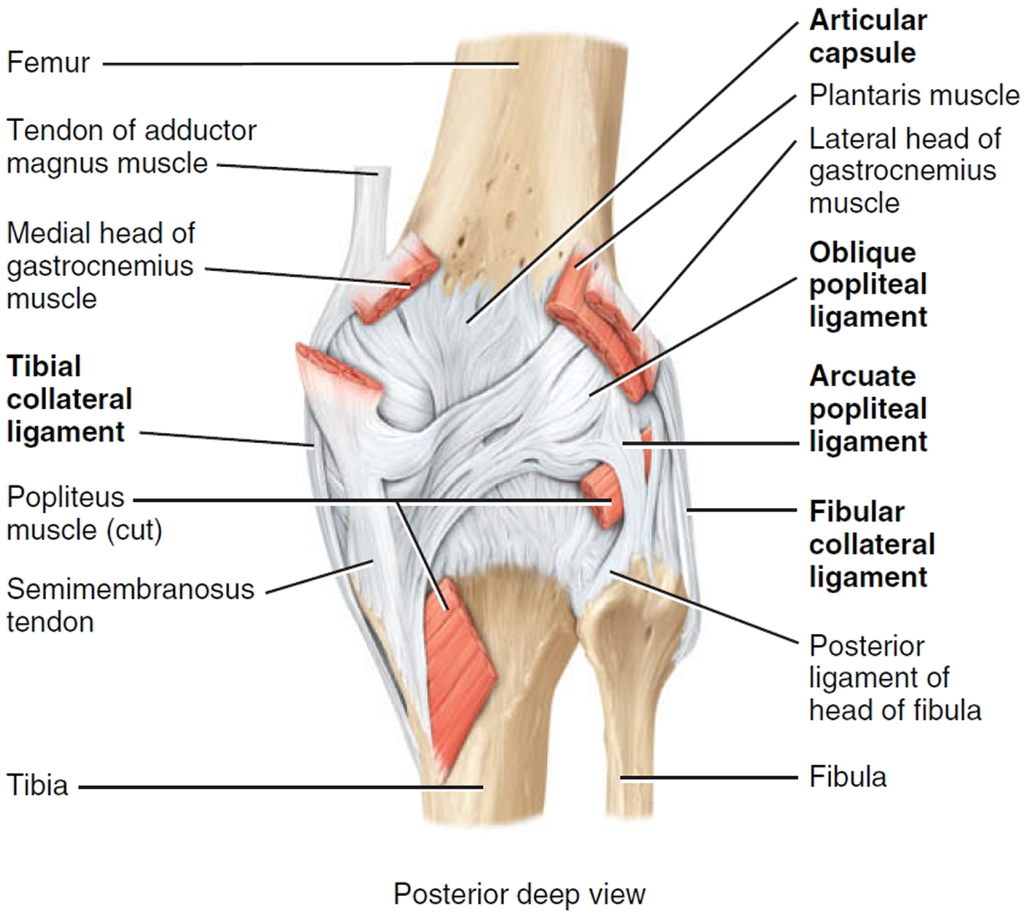
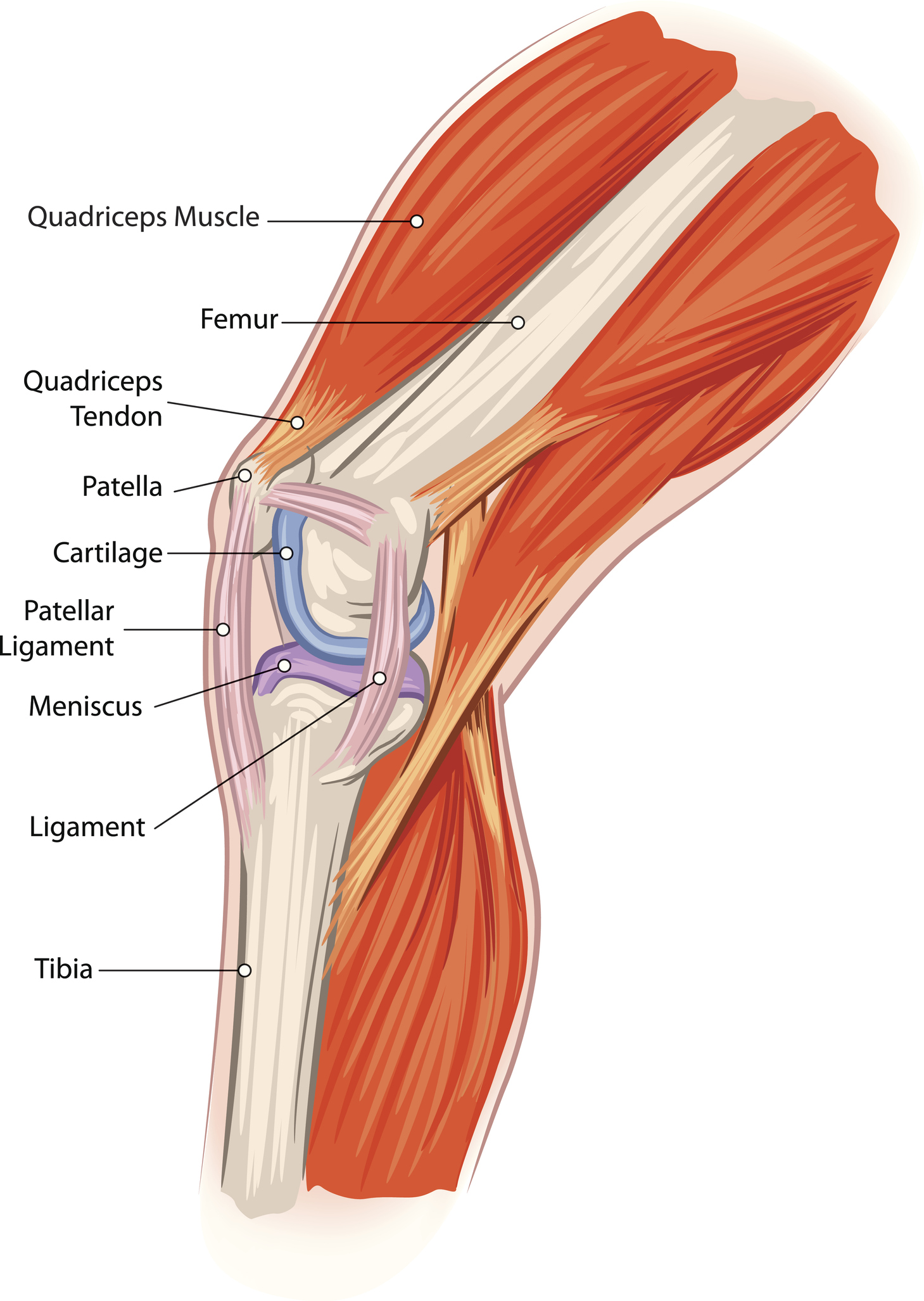
Anatomical Drawing Of The Knee - The tibiofemoral joint is an articulation between the tibia and the femur, while the patellofemoral joint is an articulation between the patella. Web to draw the knee, begin by visualizing the bones and tendons underneath to help with the placement of landmarks. It is a complex hinge joint composed of two articulations; The patella is a small, triangle shaped bone that sits. Fred flandry, md, facs*w and gabriel hommel, md* abstract: Stability of the joint is governed by a combination of static ligaments, dynamic muscular forces, meniscocapsular aponeurosis, bony topography, and joint load. Stabilizing you and helping keep your balance. This fluid is known as the synovial fluid. Finally, draw in the hamstrings covering the. Then draw the quadriceps muscles, and indicate the patella and its tendon down to the lower leg. The muscles that affect the knee’s movement run along the thigh and calf. Supporting your body when you stand and move. Fred flandry, md, facs*w and gabriel hommel, md* abstract: The structure of a normal knee joint. Stabilizing you and helping keep your balance. The knee joint is the largest joint in the human body. This fluid is known as the synovial fluid. Web the main features of the knee anatomy include bones, cartilages, ligaments, tendons and muscles. Web to draw the knee, begin by visualizing the bones and tendons underneath to help with the placement of landmarks. They are attached to the femur. Knee anatomy for figurative artists. Finally, draw in the hamstrings covering the. The knee joint is a synovial joint this means it contains a fluid that lubricates it. They are attached to the femur (thighbone), tibia (shinbone), and fibula (calf bone) by. Illustration of the human knee joint anatomy. The knee is the joint in the middle of your leg. What does the knee joint do? Web the femur (thigh bone), tibia (shin bone), and patella (kneecap) make up the bones of the knee. Functionally, the knee comprises 2 articulations—the patellofemoral and tibiofemoral. Web this article takes a concise look at the anatomy of the knee joint and describes. Web this article takes a concise look at the anatomy of the knee joint and describes the processes and conditions that cause pain in the different aspects (parts) of the knee. The knee is the joint in the middle of your leg. All these parts combine and work together. The largest joint in the body, the knee is also one. Cartoon illustration of the human knee joint anatomy. Web the knee joint is a synovial joint that connects three bones; There are four knee bones that fit together to make two different knee joints: The largest joint in the body, the knee is also one of the most easily injured. Knee anatomy involves more than just muscles and bones. Cartoon illustration of the human knee joint anatomy. Web anatomy of the knee. They are attached to the femur (thighbone), tibia (shinbone), and fibula (calf bone) by. Web to draw the knee, begin by visualizing the bones and tendons underneath to help with the placement of landmarks. Supporting your body when you stand and move. Web the knee joint is a synovial joint which connects the femur (thigh bone), the longest bone in the body, to the tibia (shin bone). Knee anatomy for figurative artists. Stability of the joint is governed by a combination of static ligaments, dynamic muscular forces, meniscocapsular aponeurosis, bony topography, and joint load. The knee is a complex joint that flexes,. Damage in even one part can hinder the functioning of the knee. Web anatomy of the knee. The tibiofemoral joint and patellofemoral joint. Then draw the quadriceps muscles, and indicate the patella and its tendon down to the lower leg. Web normal anatomy and biomechanics of the knee. Web explore innerbody's 3d anatomical model of the knee joint, one of the most important joints in the human body. The structure of a normal knee joint. Ligaments, tendons, and cartilage work together to connect the thigh bone, shin bone, and knee cap and allow the leg to bend back and forth like a hinge. Web the anatomy of the. Web the knee joint is a synovial joint which connects the femur (thigh bone), the longest bone in the body, to the tibia (shin bone). Cartoon illustration of the human knee joint anatomy. Then draw the quadriceps muscles, and indicate the patella and its tendon down to the lower leg. Web to draw the knee, begin by visualizing the bones and tendons underneath to help with the placement of landmarks. The structure of a normal knee joint. In this page, we will take a look at all of the above as well as the anatomy of the knee. Web the anatomy of the knee consists of bones, muscles, nerves, cartilages, tendons and ligaments. Illustration of the human knee joint anatomy. The largest joint in the body, the knee is also one of the most easily injured. The femur, tibia and patella. This fluid is known as the synovial fluid. Knee anatomy for figurative artists. The ligaments provide stability during loading while the muscles around the knee have a secondary role in stabilising this joint. I am sure you have seen paintings and drawings by the old masters which depict the knee area so realistically that you have no doubts about its shape. The knee is the joint in the middle of your leg. Web the main features of the knee anatomy include bones, cartilages, ligaments, tendons and muscles.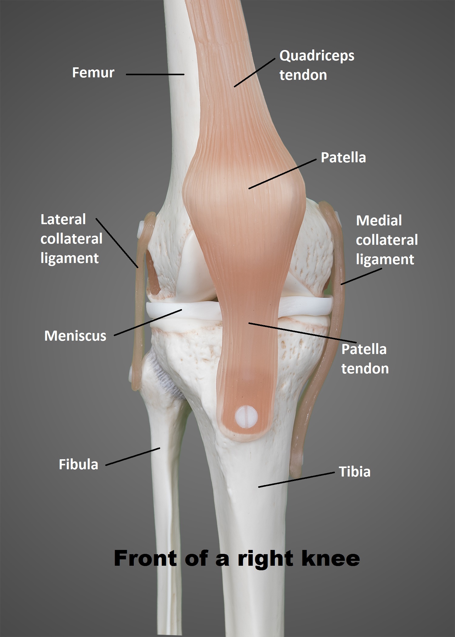
The Knee UT Health San Antonio
Knee injuries causes, types, symptoms, knee injuries prevention & treatment
Anatomy, Pathology & Treatment of the Knee Joint Articles & Advice

Anatomy of the knee Art as Applied to Medicine

The Knee Joint Anatomy Sketch
![Runner’s Knee Causes And Treatment [𝗣]𝗥𝗲𝗵𝗮𝗯](https://i1.wp.com/theprehabguys.com/wp-content/uploads/2020/08/knee-anatomy.png?ssl=1)
Runner’s Knee Causes And Treatment [𝗣]𝗥𝗲𝗵𝗮𝗯

Anatomy of Knee
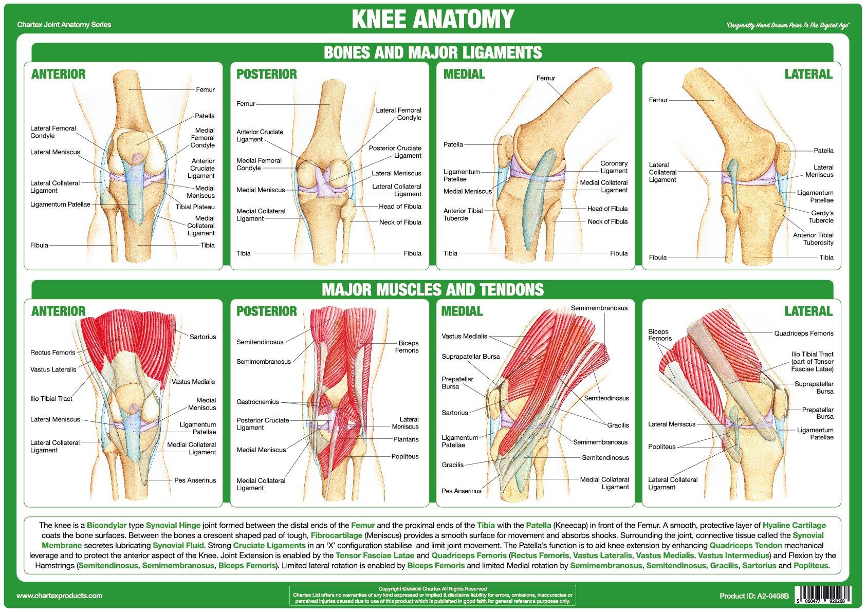
Knee Joint Anatomy Poster
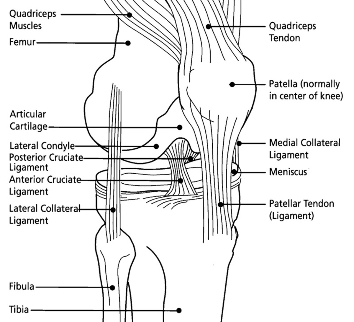
Anatomy of the Knee Joint (With Diagrams and XRay) Owlcation

FileKnee diagram.svg Wikipedia
Ligaments, Tendons, And Cartilage Work Together To Connect The Thigh Bone, Shin Bone, And Knee Cap And Allow The Leg To Bend Back And Forth Like A Hinge.
Stabilizing You And Helping Keep Your Balance.
Front View Of Normal Knee Joint.
Web The Knee Joint Is The Largest Joint In The Body And Connects The Thigh With The Lower Leg.
Related Post: